You'll find here information on 2-D gel electrophoresis and on the DiGE technology, and a description of the equipment avalable at CIAN.
Click on tabs below for information!
2-D Gel Electrophorsis and DiGE Technology
What is 2-D gel electrophoresis?
Two-dimensional protein gel electrophoresis (2-D) is a technique for separating the components of a complex protein mixture on an acrylamide gel in two dimensions, based on isoelectric point (pI) and molecular weight (MW). Depending on the sample complexity, a 2-D gel can resolve as much as 10-fold more polypeptides than an equivalent SDS PAGE gel. 2-D PAGE analysis provides a high-resolution map of protein complexity (e.g. isoforms, splice variants), post-translational modifications (PTMs) and relative abundance.
What is DiGE?
Difference Gel Electrophoresis (DiGE) is a modification of 2-D PAGE. Two or three separate protein samples are labeled with different fluorescent dyes prior to separation, enabling accurate analysis of differences in protein abundance between samples.
We use CyDye TM DIGE Fluor dyes (Cy2, Cy3, and Cy5). These dyes show the following characteristics:
| Size- and charge matched | The same labeled protein from different samples will migrate to the same position, regardless of the dye used |
| pH insensitive | No change in signal over the wide pH range used during first-dimension separation (IEF) and equivalent migration in SDS gels |
| Spectrally resolvable | The distinct signal from each fluor contributes to the accuracy |
| Highly sensitive and bright | As little as 125 pg of protein can be detected |
| Photostable | There is minimal loss of signal during labeling, separation, and scanning |
The advantages of DiGE over traditional 2-D
| Multiplexing | The labeled samples are mixed and then separated on the same 2-D PAGE gel. Thus, for samples on the same DiGE gel, gel-to-gel variation is completely eliminated, and the number of gels needed for one experiment can be cut two- to three-fold. |
| Gel-to-gel comparison | One of the three samples on a DiGE gel can be a mixture of equal amounts of all experimental samples, a “pooled internal standard”. This creates a standard for each protein in the analysis. Therefore, comparisons across different gels can be made with a high degree of confidence |
| User-friendly manipulation | Large format gels are cumbersome to handle. Since in DiGE the proteins are pre-labeled, DiGE gels do not have to be manipulated after electrophoresis. Additionally, the scanner that is used for imaging accepts gel sandwiches including the glass plates. This further reduces the variation between gels, and the risk of damaging or destroying gels |
The advantages of DiGE at CIAN
| Reagent provided | All you bring is your samples |
| Cost-effective | As a platform, we order large amount of dyes and get more interesting discount than individual lab could get, ensuring users to have the lowest fees possible |
| Support | Training is ensured by our DiGE coordinator |
| Analysis tool available | Analysis can be done on site using Decyder |
2-D/DiGE and Fluorescence Scanning Equipment
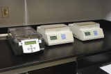 | 1st dimension:IEF: IPG-PhorII (GE)Protean IEF (BioRad) |
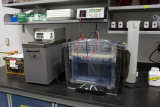 | 2nd dimension:GE Ruby SE600 (16x16cm)GE Ettan DALTsix (26x20cm) BioRad Criterion pre-cast (13x9cm) BioRad Protean (8.6x7cm) |
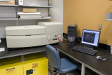 | Fluorescence Scanner:GE Typhoon Trio+ Scanner Analysis software:GE DeCyder for DiGE gels |