The History of Electron Microscopy
https://authors.library.caltech.edu/5456/1/hrst.mit.edu/hrs/materials/public/ElecMicr.htm
History of the Electron Microscope in Cell Biology
http://www.fen.bilkent.edu.tr/~physics/news/masters/ELS_HistoryEM.pdf
The First American Electron Microscopes (from the Microscopy Society of America)
An electrostatic emission electron microscope, using a standard cathode ray tube, was demonstrated in 1934 at the University of Toronto by Walter Kohl, a visiting lecturer from Germany. It was likely the first example of electron microscopy of any kind in North America.
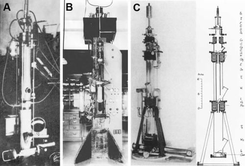
The first transmission EM constructed outside Germany was completed in 1935 or 1936 by Paul Anderson and Kenneth Fitzsimmons of Washington State University (Fig. 1A). Gordon H. Scott at the Medical School of Washington University in St. Louis started an EM program in the same year with an emission EM in 1935 and a transmission EM in 1939, constructed by Sterling Newberry (Fig. 1B). Neither of these programs continued. The resolution did not exceed that of the light microscope.
Also, in 1935, E.F. Burton established an EM program at the University of Toronto, starting with the first instrument constructed by Cecil Hall, followed by the construction by James Hillier and Albert E. Prebus 1938 of the first high-resolution EM in North America (Fig. 1C).
Hillier and Prebus were succeeded by William Ladd and John H.L. Watson, who refined the EM to yield a resolution of better than 10 nm in 1939, matching the performance of the German EMs at the time.
Hillier was hired by Alexander Zworykin (a pioneer in television development) and designed highly successful electron microscopes for RCA, eventually becoming RCA's overall head of research.
The Development of the Electron Microscope in Canada
History of the Electron Microscopy (EM) Laboratory in the Department of Anatomy & Cell Biology (formerly Department of Anatomy)
Gary C. Bennett and S Kelly Sears
The history of electron microscopy in the Department of Anatomy is principally associated with Prof. Charles P. Leblond and Prof. Yves Clermont, pioneers in biological electron microscopy. It begins in the 1940s with purchasing the RCA EMU-2B transmission electron microscope (TEM; Fig. 1a and b). For the first few years, this instrument was housed not in the department but in the Department of Physics in the Eaton Electronics Building. In 1958, the first electron microscope in the department was installed in the EM Laboratory in the Strathcona Anatomy & Dentistry Building (formerly Strathcona Medical Building [see also the history of the SADB]; Fig. 2) was a Siemens Elmiscope 1 TEM. The Elmiscope 1 was used heavily until 1972 and sold to the Montreal General Hospital.
(a)  (b )
(b ) 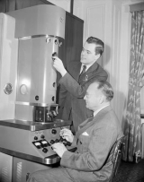
Figures 1a and b. RCA EMU-2B TEM (currently on display at the Strathcona Anatomy & Dentistry Building).

Figure 2. Siemens Elmiskop 1 TEM.
A Siemens Elmiscope 1A 100 kV TEM, was purchased in 1964 from Université Laval. Despite its bad reputation, the 1A was used for 21 years and was one of the longest-serving microscopes in the department (Fig. 2).
(a) 
(b) 

Figures 2a and b. (a) Siemens Elmiskop 1A 100 kV TEM. See also <http://physicsmuseum.uq.edu.au/elmiskop-electron-microscope>. (b) Dr. R. I. Birks shows a new Siemens Elmiskop 1A transmission electron microscope to Sir Henry Dale, National Institute of Medical Research, London, and Professor' Hank' Frank Campbell MacIntosh, Department of Physiology, presented to McGill University circa 1961. "In Canada, home of the first practical electron microscope, the Elmiskop 1 found a particularly receptive market. By 1963, several universities and labs had purchased the microscope, including McGill University (which had three, two in the Department of Anatomy), the University of Alberta, the National Research Council of Canada, and Atomic Energy Canada Limited."
Soon after obtaining the Elmiscope 1a, the RCA EMU-2B TEM was moved from the Eaton Electronics Research Laboratory to the departmental EM Laboratory and was operational until 1967. The RCA EMU-2B TEM is currently displayed in the basement of the Strathcona Anatomy & Dentistry Building. In the mid-1960s, the department obtained two Hitachi HS-7S 80 kV TEMs (Fig. 3). These served for almost 15 years before selling one to the Royal Victoria Hospital and the second to an art restorer. In 1972, Siemens Company offered the department a demonstrator model of the Elmiskop 101 TEM purchased for $25,000 (Fig. 4). This microscope was in service for many years and trained all new researchers in the laboratory.


Figure 3. Hitachi HS-7S 80 kV TEM Figure 4. Siemens Elmiskop 101
A new generation of instruments came online in the late 1970s and early 1980s. The moving parts in electron microscopes were replaced with integrated circuits in the new age of computers. With help from the Faculty of Medicine, the department acquired two new TEMs, the Philips EM400 and Philips EM400T (Figs. 5 and 6). The EM400 was the workhorse for the department until 2000 when it was replaced by the Philips EM410 TEM donated by the Department of Biology. The EM400T had a double condenser aperture system optimized for high-resolution materials research but did not provide sufficient contrast for imaging biological samples. Fortunately, the department was given a previously owned Philips EM301 TEM, which supplanted the EM400T until both were decommissioned in early 2000 (Fig. 7).
![]()
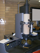
Figure 5. Philips EM400 120 kV TEM Figure 6. Philips EM400T 120 kV TEM
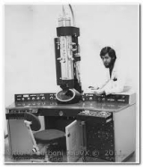

Figure 7. Philips EM301 100 kV TEM
In 1986, the department received a $600,000 MRC Major Equipment Grant to purchase the JEOL JEM-2000FX S/TEM equipped with Kevex X-ray energy dispersive spectrometer (XEDS). This instrument also was equipped for scanning-transmission electron microscopy (S/TEM; Fig. 8). Unlike a regular SEM that can detect secondary electrons generated from the surface of a specimen, in S/TEM mode, the rastered excitation electron beam passes through the thin biological section, generating secondary electrons that are detected and analyzed. The XEDS system in this microscope enabled the identification and localization of virtually any element in biological tissues. Prof. Hershey Warshawsky used it to detect calcium and iron atoms in the enamel sections of teeth. On the occasion of the inauguration of this "state-of-the-art" instrument, Warshawsky published a brief history of the departmental EM Laboratory in the McGill Reporter (Feb 13, 1986).
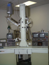
Figure 8. JEOL JEM-2000FX 200 kV S/TEM
Scientific and technical staff: The EM Laboratory was administered initially by Dieter Curlis, the chief technician. He was succeeded by Ed Sanborn and his technician, Pat Coen. After leaving McGill for Université de Montréal, Sanborn was succeeded by Bertie Van Heyningen. Van Heyningen was followed by Prof. Hershey Warshawsky, the first long-term director of the EM Laboratory (1965 to 1973). Warshawsky played a crucial role in obtaining many of the departmental electron microscopes. Prof. Michael Lalli succeeded Warshawsky as director of the EM Laboratory and served until 1998, except for one year when Prof. Louis Hermo was director. Other technical staff who worked in the lab over the years included Julius Batky (founder of Marivac/Canemco, a Canadian supplier of EM equipment and consumables), Ingrid Simons, Henry Szczawinzki, Kathy Hewitt, Ruth Partridge, Cathy Tang, Sonia Bujold, Aliki Michaelidou, Patricia Hales, Matilda Cheung, Caroline Tanguay, Jeannie Mui, Lee Ann Monaghan, and many others.
History of the Facility for Electron Microscopy Research (FEMR)
Critical investments and commitments: It was evident by the late 1990s that acquiring, operating, and maintaining expensive electron microscopes and ancillary equipment with users widely distributed across faculties and departments on the downtown campus of McGill was a severe organizational challenge. Instruments and equipment were used primarily within a single department and therefore were individually responsible for identifying and securing appropriate funding sources for operation and maintenance. Such organization resulted in duplication of instruments, services, technical and scientific personnel, and, more importantly, increased costs. As a long-term solution to chronic underfunding, it was proposed to create a core facility for electron microscopy at McGill with shared instrumentation that would greatly benefit multi-faculty and multi-departmental users of expensive scientific instruments.
Under the leadership of Prof. Hojatollah Vali as Director and Dr. S Kelly Sears as Research Manager, the FEMR was formally launched in 2001 with functional but obsolete research infrastructure. The grouping of many of the electron microscopy (EM) laboratories into a single cross-departmental, cross-faculty and cross-institutional core facility was seen as a mechanism for (i) consolidating the strengths and rectifying weaknesses in imaging, (ii) enriching and expanding cross-disciplinary and interdisciplinary interaction and research in medicine, science, and engineering, (iii) improving the quality and expanding the scope of research and research-training by providing shared instrumentation that fosters the integration of research and education in research-intensive learning environments, and (iv) assuring the financial stability of research and service delivery and technological enhancement. This was viewed as especially relevant as advanced research instruments that were once of interest only to specialists are increasingly required by many scientists to solve critical research problems. Researchers who depend on advanced instruments require highly specialized knowledge and expert training in proper operation and use. Moreover, the committed sharing of advanced infrastructure to the research community offered the potential to maintain affordable user fees. At the same time, technology‐based research activities and technology development, including significant equipment purchases, continues to be investigator‐driven through research, equipment, and maintenance grants.
Since its inception, the FEMR has worked with researchers from all academic institutions in Montreal and elsewhere in Quebec to acquire the expertise and cutting-edge infrastructure to ensure researchers have the tools to conduct world-class research. The FEMR continues to be investigator-driven through instrument and operation/maintenance grants for technology-based research activities and technology development, including significant equipment purchases. The FEMR has thus evolved through generous government and institutional support, including three CFI infrastructure awards (CFI-LEF 744, CFI-LEF 11505, and CFI-LEF 30797) and numerous NSERC and CIHR operations and maintenance and equipment grants, and the cooperation of researchers from institutions throughout the region of Montreal, into a world-class multi-user, multidisciplinary, multi-faculty, multi-institutional shared equipment facility. With state-of-the-art light and electron microscopy research infrastructure, the FEMR operates on a cost-recovery basis. In 2000, a CFI-LEF 744 (PI - H. Vali, $8.9 million) grant created the Montreal Network for Materials, Molecular and Structural Imaging (MNMSMI), a regional centre providing facilities and a multidisciplinary environment to pursue world-class science, to develop world-class technologies, and to maintain outstanding capabilities in electron microscopy research. The MNMSMI was designed as a consortium led by the FEMR in partnership with the Laboratoire de Recherche sur les Tissus Calcifiés et Biomatériaux (Université de Montréal) and the Centre de Caractérisation Microscopique des Matériaux (CM)2 (École Polytechnique) (See Table 1 for a list of instruments and equipment).
Table 1. Major Instruments & Equipment obtained from CFI-LEF 744.
| Instrument/Equipment | Location |
|---|---|
| Hitachi S-4700 FE-SEM | McGill University |
| Hitachi S-3000N VP-SEM | McGill University |
| JEOL JSM-6340F FE-SEM | Université de Montréal |
| JEOL JEM-2100F 200 kV FE-TEM | École Polytechnique |
| Hitachi FB-2000A FIB | École Polytechnique |
| Gatan Multiscan CCD Camera Model 791 (x2) | McGill University/Université de Montréal |
| Leica Microsystems EM UCT Ultramicrotome | Université de Montréal |
| Leica Microsystems EM BAF060 Freeze-Fracture System | McGill University |
In 2008, a CFI-LEF grant (11505; PI - J. Bergeron) was awarded to the FEMR as the lead institute and to Université Laval to create the regional Facility for Electron Cryomicroscopy and Cryo-Electron Microscopy (FECCET). A total of $8.03M, combined with $736K from a CFI-LOF Grant (12824) awarded to Dr. I. Rouiller, a recruit in structural biology and cryo-EM at the FEMR and appointed in the Department of Anatomy & Cell Biology, enabled the acquisition of state-of-the-art transmission electron microscopes and ancillary equipment for advanced sample preparation (Table 2).
Table 2. Major Instruments & Equipment obtained from CFI-LEF 11505.
Equipment Location
| Instrument/Equipment | Location |
|---|---|
| FEI Titan Krios 300 kV Cryo-STEM w/Gatan Imaging Filter | McGill University, Université Laval |
| FEI Tecnai G2 F20 200 kV Cryo-STEM | McGill University, Université Laval |
| FEI Tecnai G2 Spirit 120 kV Cryo-TEM | McGill University, Université Laval |
| FEI Vitrobot Mark IV (x2) | McGill University, Université Laval |
| Leica Microsystems EM PACT2 High-Pressure Freezer (x2) | McGill University / Université Laval |
| Leica Microsystems EM AFS2 Automatic Freeze Substitution | McGill University |
| Leica Microsystems EM CPD030 Critical-Point Dryer | McGill University |
| Leica Microsystems EM UC7/FC7 Cryo-ultramicrotome | Université Laval |
| High-Performance Computing | McGill University /Université Laval |
In 2012, a CFI-LEF grant (30979; PI - M. McKee) of $4.54 million was awarded to the FEMR to advance the multi-modal workflow capability for cryogenic correlative light-electron microscopy (cryo-CLEM; Table 3).
Table 3. Instruments & equipment obtained from CFI-LEF 30797.
Equipment Location
| Instrument/Equipment | Location |
|---|---|
| FEI Helios Nanolab 660 DualBeam Leica Microsystems EM VCT100 Cryogenic Transfer System | McGill University |
| FEI CorrSight Digital Fluorescent Microscope | McGill University |
| Leica Microsystems EM UC7/FC7 Cryo-ultramicrotome | McGill University |
| Leica Microsystems EM ACE600 High-Resolution Sputter Coater | McGill University |
| High-Performance Computing | McGill University/CalCul Quebec/Compute Canada |
Due to the CFI investments over this period, the FEMR has been highly successful in obtaining almost $10M worth of peer-reviewed, tri-council operation and maintenance grants and other equipment and support grants (Table 4). In addition, as cutting-edge research increasingly relies on the services and specialized equipment that is too costly for departmental or individual procurement, many researchers across disciplines and faculties have acquired funding for their research projects based on the availability of state-of-the-art infrastructure and expertise at the FEMR.
Table 4. Equipment, Operation & Maintenance Grants 1999-2018.
| Funding Agency/ Program | PI. | Purpose | Amount | Year(s) Awarded |
|---|---|---|---|---|
| CIHR MME (MT- 14210) |
Bergeron, J. | JEOL JEM-2011 TEM, service contracts, & salary support | $703,878 | 1999-2002 |
| McGill University | Vali, H. | Cash contribution to FEMR | $1.16 Million | 2001-2014 |
| McGill University | Bergeron, J. | Service contracts; salary support | $670,530 | 2002-2007 |
| FQRNT-CBB | Tabrizian, M. | Salary support | $550,000 | 2002-2011 |
| McGill University | Vali, H. | Renovations to Rooms B/4 to B/9, B/21 to B/27 | $275,000 | 2003 |
| CIHR-MME (MME- 67417) |
Bergeron, J. | FEI Tecnai 12 120 kV TEM & Leica Microsystems UCT Cryo-ultramicrotome | $295,884 | 2003 |
| McGill University | Vali, H. | Quartz X1 EDS on JEOL JEM-2000FX TEM | $36,000 | 2004 |
| NSERC-RTI | Gauvin, R. | STEM detector on Hitachi S-4700 FE-SEM and EBSD on Hitachi S-3000N VP-SEM | $146,000 | 2007 |
| CIHR-RRG (MT- 165625) |
Bergeron, J. | Service contracts; salary support | $689,280 | 2007-2012 |
| McGill University | Vali, H. | AMT XR-60B CCD Camera and EDAX Genesis EDS on Philips CM200 TEM | $80,000 | 2008 |
| CFI-IOF (11505) | Bergeron, J. | Operational Support | $963,6635 | 2008-2014 |
| CFI-LOF (12824) | Rouiller, I. | Upgrade of FEI Tecnai G2 F20 Cryo-TEM | $736,153 | 2008-2014 |
| FRSQ-GRASP | Gehring, K. | Salary support | $80,000 | 2009-2012 |
| CFI-LOF | Gauvin, R. | SU8000 FE-SEM with an EDAX SDD EDS and EBSD system | $904,000 | 2009 |
| CFI-LEF | McKee, M. | Gatan Precision Ion Polisher (PIPS) | $70,000 | $70,000 2009 |
| McGill University | Vali, H. | Renovations Rooms B/21 to B/27; B/31, B/32, B/32A, B/32B | $4.5 Million | 2009-2010 |
| NSERC-RTI (EQPEQ 406251-11) |
Rouiller, I. | AMT XR-80C CCD camera for FEI Tecnai G2 Spirit Cryo-TEM | $83,122 | 2011 |
| NanoQuébec - MCRF | Tabrizian, M. | Salary support | $320,000 | 2011-2014 |
| McGill University | Vali, H. | Leica Microsystems Critical Point Dryer EM CPD030 | $20,000 | 2012 |
| CFI-LOF | Siwick, B. | Gatan Ultrascan 1000 CCD Camera for Philips CM200 TEM | $100,000 | 2012 |
| FRQ-S-GRASP | Gehring, K. | Salary support | $100,000 | 2013-2018 |
| CFI-IOF (30797) | McKee, M. | Operational support | $400,000 | 2013-2018 |
| McGill University | Gauvin, R. | Hitachi SU8230 FE-STEM | $550,000 | 2013 |
| FEI Company | Vali, H. | Operational and maintenance support | $650,000 | 2013-2018 |
| McGill University | Vali, H. | FEI Quanta 450 FE-ESEM & FEI Inspect F-50 FE-SEM | $750,000 | 2013 |
| NSERC-RTI | Kollman, J. | Gatan Model 626 Single Tilt Liquid Nitrogen Cryo Transfer Holder | $70,000 | 2013 |
| CFI-LOF | Tamimi, F. | iXRF X-Beam micro-X-ray fluorescence (XRF) system for FEI Inspect F-50 FE-SEM | $100,000 | 2013 |
| CFI-LOF (32601) | Schmeing, M., Rouiller, I., Kollman, J. | FEI Falcon 2 Direct Detection Device (DDD) & Tomography 4 SW on Titan Krios 300 kV Cryo-STEM | $743,200 | 2014-2019 |
| CFI-IOF (32601) | Schmeing, M., Rouiller, I., Kollman, J. | Operational support | $90,000 | 2014-2019 |
| FEI Company | Vali, H. | Phase plate on Titan Krios 300 kV Cryo-STEM | $1 million | 2014 |
| McGill University | Vali, H. | EDAX Octane Silicon Drift Detector (SDD) Microanalytical System for FEI Tecnai G2 F20 Cryo-STEM | $80,000 | 2014 |
| CFI-JELF | Moores, A. | Thermo Scientific Talos 200FX G2 TEM | $2,000,000 | 2017 |
| CFI-JELF | Ortega, J., Vargas, J., Tocheva, E. | Gatan GIF BioQuantum LS and K3 DED | $1,900,000 | 2018 |
| NSERC-RTI | Vargas, J & Strauss, M | TVIPS XF416 16MP CMOS Camera | $140,000 | 2019 |
| CFI-JELF | McKee, M. | Leica EM VCT500 Cryotransfer System, EM ACE600 Sputter Coater, and EM CPD300 CPD | $281,000 | 2019 |
| McGill University - Office of the Provost | Ortega, J. | Operational Support | $300,000 p.a. | 2020 - |
| CFI- Innovation Fund | Mountanabbir, O, McKee, M, Gauvin, R... | Leica EM ICE HPF | $350,000 | 2020 |
| CFI-Innovation Fund | Ortega, J. et al. | Multiscale 3D Cryo Imaging: From Organs to Molecules | $20,702,128 | 2023 |
Since 1999, the Office of the Vice-Principal Research & International Relations (OVPRIR) and the Faculties of Dentistry, Engineering, Medicine, and Science have provided a cash contribution of $135K yr-1 to FEMR for salaries of scientific and technical staff. In 2019, the Office of the Provost agreed to provide $300,00/year for operations.
As research space is an institutional resource, the Faculties of Dentistry, Engineering, Medicine, and Science have allocated space for FEMR based on McGill's programmatic needs and strategic research priorities. The most significant part of the assigned space for FEMR is in the basement of the Strathcona Anatomy & Dentistry Building, including its Wet Lab, Sample Preparation Lab, and five rooms for TEMs and SEMs. The FEMR also has space in the W.H. Wong Engineering Building (TEM and SEM). In terms of costs for renovation and construction, McGill invested $275,000 in 2003 to renovate the Wet Lab and Cryo-Lab in the SAD Building. In 2009, McGill invested $4.5M to renovate and update the research space to install the Titan Krios 300 kV cryo-STEM, Tecnai G2 F20 cryo-STEM, and the Tecnai G2 Spirit 120 kV cryo-TEM.