
“To (Osler) the pathological specimen, properly prepared and catalogued, was a record of disease more instructive than any textbook description could possibly be. … Each had a wonderful beauty of its own – valuable in that it demonstrated some essential bit of knowledge of disease for which it should be preserved as an individual record.”
Warthin AS
Bulletin of the International Association of Medical Museums
1926, IX: 2.
Fifty-nine specimens remain in the Maude Abbott Medical Museum Osler collection in 2023. The vast majority originated in the approximately 800 autopsies performed by Osler during his tenure at the Montreal General Hospital (MGH) from 1876 to 1884. It is not certain how many specimens derived from these he donated to the Museum; however, Abbott wrote in 1937 that “he must have added at least five hundred”.* We do know from Abbott’s writing that only about 180 remained in 1898 when she was appointed Assistant Curator of the Museum. Exactly what happened to the other 300, as well as many of the 120 “lost” after 1898, is not clear; however, there are undoubtedly a variety of causes.
It is likely that Osler himself was responsible for the loss of some material. Thomas McCrae, one of Osler's residents, wrote in 1926 that Osler “had a considerable number of specimens (250 or 300) in Baltimore, some of which came from Montreal ...”.** According to McCrae, he did not use them much in teaching, but “on some occasions ... went over them and ... remembered the particulars.” It is not known what became of these specimens, but as with Osler’s last two autopsy logbooks, they are lost and likely destroyed.
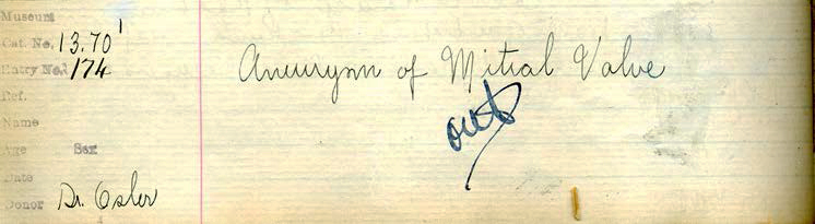
Many specimens were probably “thrown out” because of wear and tear related to student handling or deterioration in the quality of the specimen as a result of fluid discoloration or precipitate. As is evident from the current material, the string/glass rod device by which the specimens have been mounted in glass jars is quite delicate (Specimen 17). Although this mechanism of display differs from that used in the 1870s/1880s, it is likely that whatever was done by Osler or his assistants was also easily broken by the inattentive student. A review of the first Museum logbook shows 35 entries indicating Osler as donor in addition to those associated with specimens still present in 2013. As these entries indicate, some specimens were discarded for unspecified reasons; however, many others are simply unaccounted for. These “lost” specimens show a wide variety of abnormalities, some well documented in the surviving collection (such as endocarditis (3 specimens) and aortic aneurysms (2 specimens)), but others which are not represented and whose absence is a significant loss. Examples include infectious disease (such as hepatic/colonic amebiasis, hepatic hydatid disease (echinococcosis) and smallpox) and pneumoconiosis (three specimens illustrating coal miner's "anthracosis", including one probably showing progressive massive fibrosis and another the disease in a miner's dog!).
Not all specimens were lost because of student neglect or specimen deterioration. A significant number perished in the fire of 1907, which destroyed much of the Medical Museum; however, Abbott stated that most of the circulatory, respiratory and digestive collections survived the conflagration. (Surprisingly, only a single pulmonary specimen remains.) Alvin Rodin (see farther on) documented 38 specimens said by Abbott to have been in the Museum in the 1930s, most of which are now lost. It is unlikely that student use was a factor in their disappearance, since all student teaching at that time was held in the Pathological Institute, across the street from the collection. It is possible that theft was responsible, as suggested by Abbott in a 1937 article in which she stated that "there was until recently an extremely interesting specimen in the McGill collection of the heart of a calf showing many cysticerci embedded in the musculature. This valuable specimen survived the McGill fire of 1907, but unfortunately disappeared recently from the collection".***
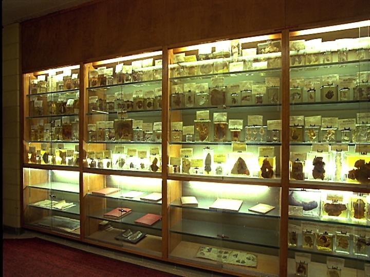
Because of the various reasons discussed above, the number of specimens decreased to about 150 in 1927, and 130 in 1934. Following Abbott's death in 1940, her museum, which had been renamed the Medical Historical Museum, was dissolved. Although details are scarce, it seems that the Osler collection was transferred to the Pathological Institute around 1945 and put in storage. The fifty-five specimens which remained in 1966 were placed in display cases in the foyer of a newly constructed wing of the Pathological Institute. Because of concerns about security and preservation in the unguarded and overheated foyer, the specimens were again moved to storage in 2002, where they remain at the time of this writing.
Four “new” specimens originating from Osler's time at the Montreal General Hospital were identified in the McGill Museum between 2002 and 2005. These are labeled “New Osler” in this exhibit. Three of them were identified in the series of 80 specimens that forms the Maude Abbott Collection of congenital heart disease (pulmonary atresia with associated ventricular septal defect and patent ductus arteriosus; a portion of atrial septum that has a thin-walled fossa ovalis; and an aortic valve with fenestrated leaflets). The fourth “new” specimen (a bicuspid aortic valve) was found mixed up with specimens that had been in storage for years in the basement of the Pathological Institute.
In addition to the moves between and within buildings, the collection has undergone several restorations over the years. The specimens were originally fixed in alcohol or Sappey’s fluid and mounted in cylindrical glass jars. Most were remounted with new supporting glass rods and string in rectangular prism jars in the 1930s (the same ones in the current collection). In 1963, the preservative fluid was replaced with Kaiserling III and the descriptive cards were retyped (giving those reproduced in this exhibit). Overall, the specimens, their glass/string supports and their preservative fluid are in remarkably good condition. However, as indicated previously, some of the glass supports have broken (See Specimen 17) and residue is evident on the tissue surface in many cases on close examination.
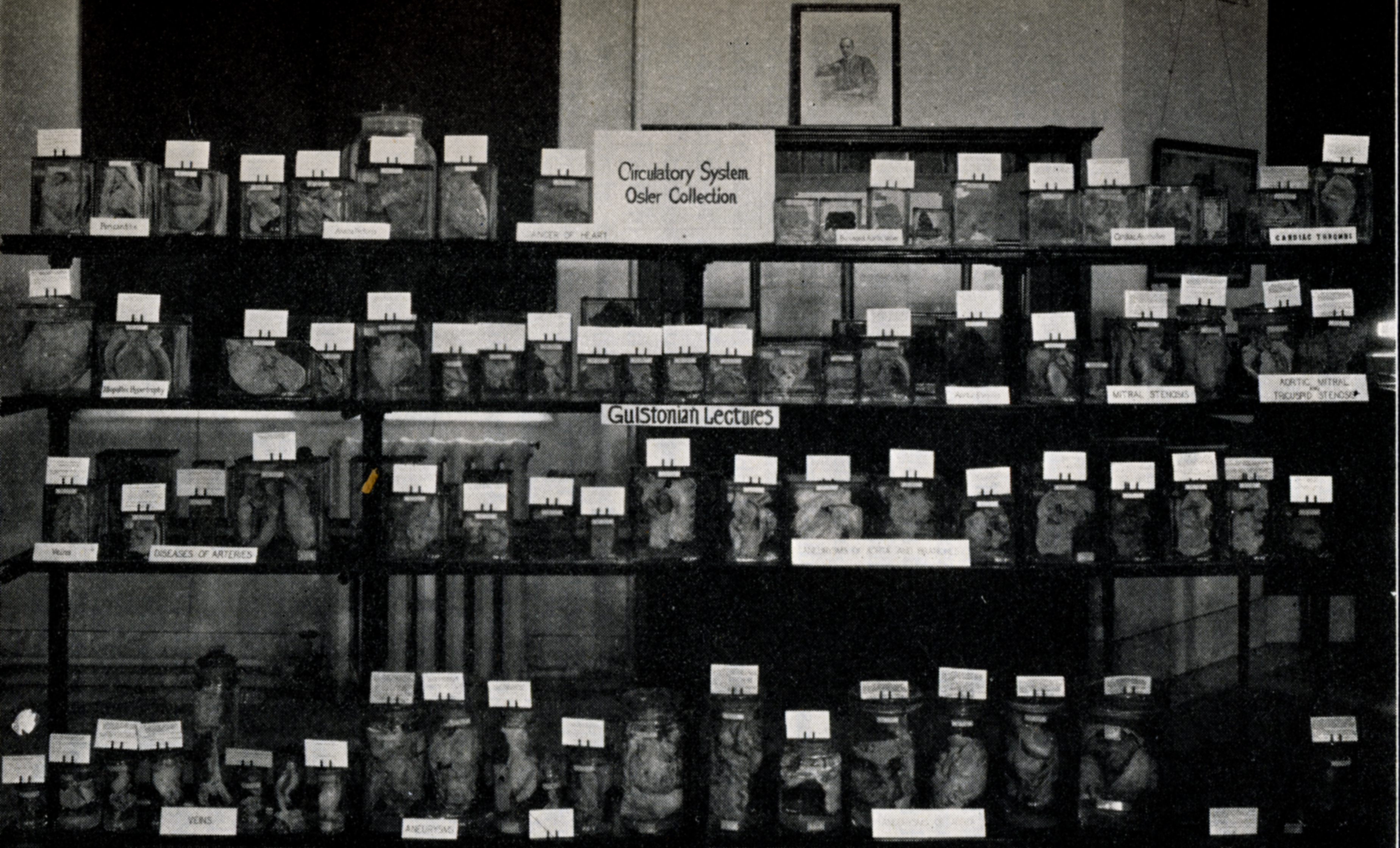
The collection has been the basis of several exhibits and the subject of a number of publications (see references below). Abbott presented the specimens at the 10th Congress of the American College of Surgeons in Montreal in 1920 and at the 27th Annual Meeting of the International Association of Medical Museums in Toronto in 1934. The 1920 display was similar in concept to that in this site, in that it included copies of Osler’s published articles which corresponded to the specimens. In the 1930s, the specimens were sequestered in Abbott's Medical Historical Museum as a permanent display.
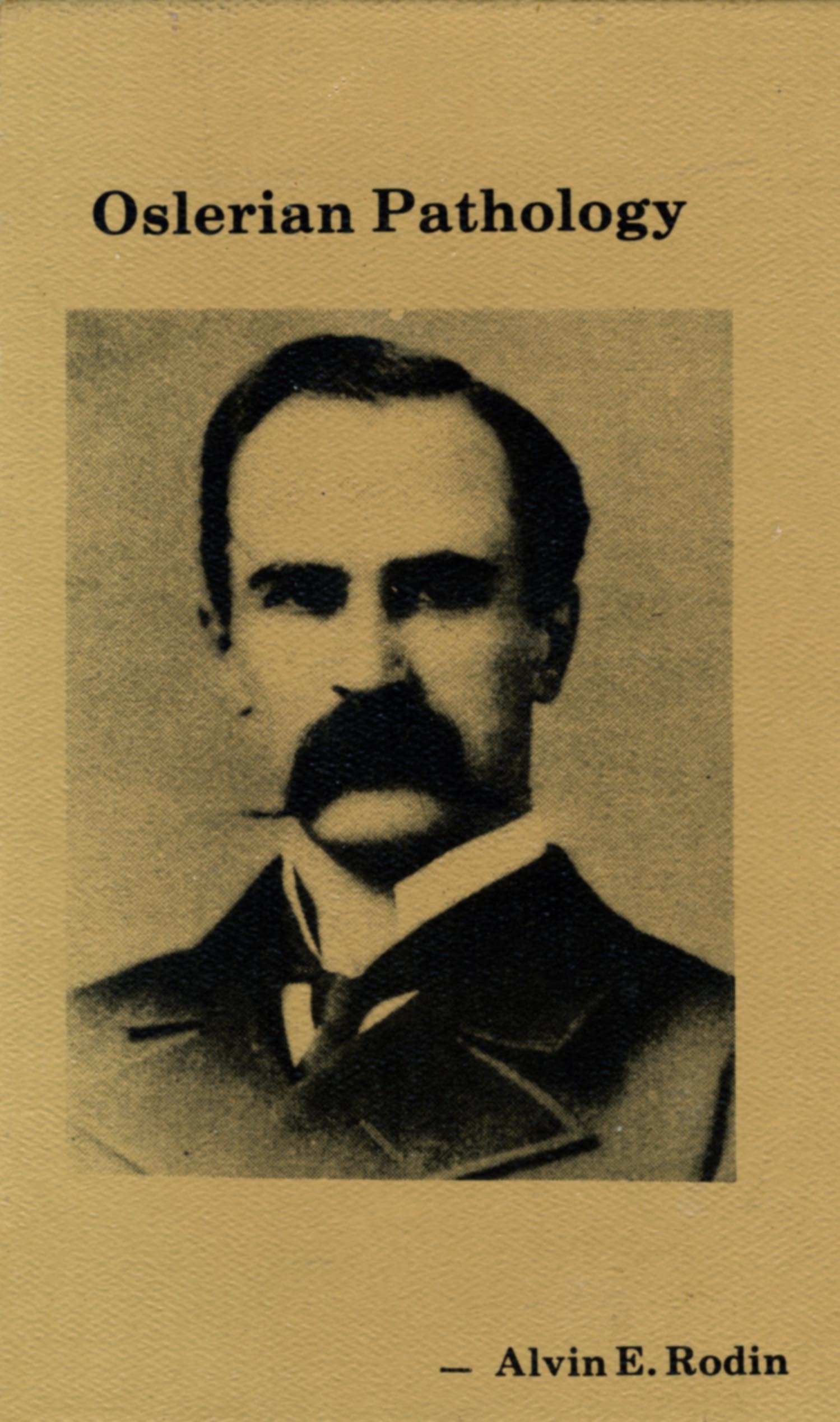
The most comprehensive publication related to the collection is a book by Alvin Rodin in 1981. In addition to black and white photographic images of 55 specimens, he included discussions of the history of the collection, of Osler’s work as a pathologist and of Osler’s concepts of the diseases illustrated by the specimens. He also transcribed Osler’s autopsy protocols which related to specific specimens; these are reproduced in this website with grateful acknowledgement of his effort.
A significant number of errors have been introduced into the collection and discussions of it over the years. Some of these may have been made because of lack of pathologist input at the time of jar restoration in the 1930s and card transcription in the 1960s. They include mounting the metal descriptive card holder on the wrong side of the specimen, resulting in the poor student having to search (possibly in vain) for the abnormality he was supposed to see (e.g., Specimen 8 and Specimen 23). Some specimens have also been incorrectly identified (as, for example, one instance in which the descriptive cards of two endocarditis specimens had been interchanged). Finally, some cards refer to disease which is not present (e.g., thickening (fibrosis) suggestive of chronic disease in what appears to be a normal valve (See Specimen 18)).
Surprisingly, Rodin (who was a pathologist) also made a number of errors in his book illustrations and the interpretation of them. Thus, some images (See Specimen 12) are mislabeled and some (See Specimen 21) purport to show an abnormality which is not present. In several cases, he notes that an abnormality indicated on the descriptive card is not evident when, in fact, it is clearly seen on the back of the specimen (See Specimen 24). We hope that most, if not all, of these errors have been identified and corrected in the review of the collection for preparation of this website.
* Abbott M. Bull Inst Hist Med 1937; 5: 765.
** McCrae T. Bulletin of the International Association of Medical Museums 1926, IX.
*** Abbott M. Bull Inst Hist Med 1937; 5: 784.
Additional Reading
Abbott M. The Osler Pathological Collection in the Medical History Museum of McGill University. Bulletin of the International Association of Medical Museums XIV March 1935 21-27.
(PDF version)
Abbott M. The pathological collections of the late Sir William Osler at McGill University. Bulletin of the International Association of Medical Museums 1926, IX: 185 – 199.
(PDF version)
Rodin A. Osler’s Museum specimens of heart disease. Their nature and significance. Chest 1971, 60: 587 – 594.
(PDF version)
Rodin, A. E. (1981). Oslerian pathology: An assessment and annotated atlas of museum specimens. Lawrence, Kansas: Coronado Press.
Link
Sir William Osler Memorial Number, Canadian Medical Association Journal (Volume 10, Special Issue, July 1920).