Interactions between the subcortical and cortical regions of the brain differ between autistic and non-autistic people, according to research published in the open access journal Nature Communications.
ACAR researcher Boris Bernhardt and his colleagues leveraged magnetic resonance imaging (MRI) data from the multi-site Autism Brain Imaging Data Exchange Initiative with innovative computer modeling techniques to study how a combination of small-scale (microcircuit) and large-scale (macroscale connectome) anomalies could explain the development of autism spectrum disorder (ASD).
Although imaging the human brain at micro and macro scales is more accessible than ever, laboratories still struggle to get large enough or varied data sets.
Open Science initiatives like the Autism Brain Imaging Data Exchange offer a solution, giving researchers unprecedented access to large-scale brain images collected by research labs around the world.
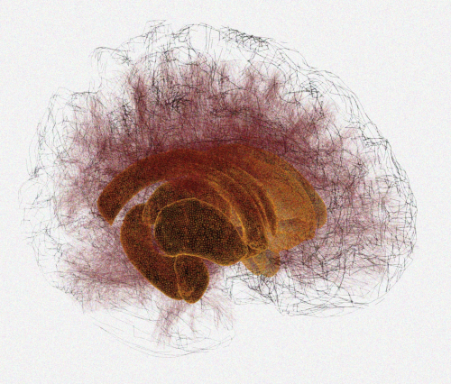
Fig. 1. Using advanced brain network analyses as well as computational modelling, the work of Park and colleagues points to an imbalanced interaction between cortical as well as subcortical networks.
To understand how the brain is wired, neuroscientists create maps, or connectomes, of neural interactions within the brain.
Connectivity explored through MRI can be determined in two ways:
- Functional: Measured by looking at what regions of the brain are generally activated together, either during a task or in resting conditions, and
- Structural: Determined by approximating the physical connections of brain areas.
A large number of studies have observed atypical brain functional networks in the brains of autistic people using fMRI, but there still is little known about the differences in structural connectivity.
Machine-learning algorithm and computational simulations – a window into the brain
For this analysis, Bernhardt and colleagues applied a machine-learning algorithm called “manifold learning” to get a clearer picture of structural connectivity alterations in the brains of 47 autistic people and 37 non-autistic people.
They also used computational simulations, to predict functional connectivity from structural connectivity and to interrogate small-scale brain circuit properties.
Differences between autistic and non-autistic groups
In the macroscale, researchers found differences between the autistic and non-autistic groups in cortical areas related to sensory and motor processing as well as in regions involved in more integrative functions, such as those in the mid-parietal cortex.
Beyond pointing to atypical cortical reorganization, the authors also observed differences in how subcortical brain areas, such as the thalamus, communicate with microcircuits in these cortical areas.
They then used gene expression data from the Allen Institute for Brain Sciences, to explore the possible neurobiological underpinnings of these connectivity findings.
Their analysis could demonstrate that the affected regions harbor genes expressed in cortical and thalamic areas in early childhood and adolescence, as well as early infancy and young adulthood.
This study highlights that the way that subcortical brain regions interact with cortical regions likely plays an important role in whole-brain connectivity anomalies in autism.
The overall disorganization of circuits in the brains of autistic versus non-autistic people likely reflects an early imbalance in the maturation of structural and functional connections in both sensory and higher order pathways, and can help to better understand the broad range of autism symptoms.
Moving forward
It will be important to increase the sample size by making a larger number of high-quality functional and structural brain scans available, and to assess younger cohorts longitudinally to trace atypical brain development directly.
As a next step, it will also be important to study individuals with a range of neurodevelopmental disorders to see how specific these imbalances are to autism.
Source: Park, By., Hong, SJ., Valk, S.L. et al. Differences in subcortico-cortical interactions identified from connectome and microcircuit models in autism. Nat Commun 12, 2225 (2021). https://doi.org/10.1038/s41467-021-21732-0
About ACAR
The Neuro’s Azrieli Centre for Autism Research (ACAR) transforms research, training and care to improve the quality of life of autistic people and their families. Established in 2017 thanks to the Azrieli Foundation, ACAR operates in the spirit of Open Science, inclusion and community collaboration. The state-of-the-art research centre is committed to advancing understanding of the mechanisms underlying autism and related conditions, developing new diagnostic tools and effective interventions through translational research and care, and training the next generation of fundamental and clinical autism researchers.