Preclinical Research PET
Preclinical PET imaging with our MicroPET system is an invaluable tool in research due to its unprecedented sensitivity, tissue penetration depth, non-invasiveness and precise dynamic quantification of biological processes in vivo. Similar to its big brother human PET, microPET has become an especially powerful tool in neuroscience research for the imaging of metabolic dysfunction, aberrant expression of receptors, enzymes and misfolded proteins in the living animal brain, used as a model for the human brain. Several exciting projects taking advantage of the animal PET scanner at the BIC include preliminary evaluation of the novel PET tracers, studies of the animal models of neurodegenerative diseases as well as testing experimental intervention procedures that have potential to be translated to humans.
About
The microPET R4 is a preclinical PET scanner developed and commercialized by CTI Concorde Microsystems/Siemens. It is made of 4 rings of 24 detector blocks, each block consisting of 8 x 8 Lutetium Orthosilicate (LSO) crystals of size 2.1 x 2.1 x 10 mm3. This design produces an axial and transaxial field of view (FoV) of 7.8 cm and 10.0 cm respectively, and with a bore diameter of 12.0 cm this camera is particularly suitable for rodent studies. With a sensitivity of 2% and a spatial resolution of 1.8 mm at the centre of the FoV, the microPET R4 can acquire, using small doses of radioactivity, dynamic PET studies of small structures of the brain with high temporal sampling.
Data is acquired in list mode, which allows post acquisition temporal framing for the reconstruction of the dynamic studies. The attenuation map is obtained with a fast acquisition in transmission using a Co-57 source. All standard corrections for image degradation factors, such as random events, attenuation, scatter, detector efficiency non-uniformity, are implemented with the iterative image reconstruction algorithms.
The most commonly studied species in microPET imaging at the Neuro are rats, both wild-type and genetically or therapeutically modified to recapitulate different aspects of human brain function in health and disease. However, recently we embarked on a new and exciting project dedicated to imaging of a small monkey species, the marmoset. As primates, these animals are better suited as translational models of the human brain than rodents.
Selected completed and ongoing microPET imaging projects at the BIC
Characterization of animal models of Alzheimer’s disease (AD) using PET biomarkers (Dr. Rosa-Neto).
Dr Rosa-Neto’s lab recently developed a transgenic (Tg) rat model, called TgAD344-AD, which recapitulates the pathophysiology of AD. Using the microPET facility at the BIC, his group follows up the pathophysiology progression in these transgenic rats with PET tracers such as [18F]FDG (hypometabolism), [18F]NAV4694 (amyloid plaques), [18F]MK6240 (neurofibrillary tangles) and [11C]PBR28 (activated microglia). He also measures tau propagation in these animals over time to study its prion-like properties. Finally, he recently started a study of sleep modulation and its effect on synapses using the novel synaptic PET tracer [18F]SDM-8. Such microPET studies in animal models will help us better understand the underlying pathophysiology of AD.
Development of novel PET tracers for unexplored neuroreceptor targets (Dr. Alexey Kostikov).
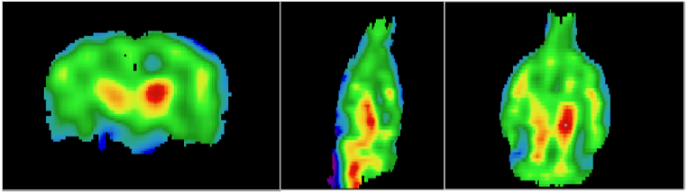
Dr Kostikov and his team is developing PET tracers for imaging of receptors interacting with neurotrophin factors, such as nerve growth factor NGF, brain-derived neurotrophic factor BDNF, NT3 and NT4, which are aberrantly expressed in the early stages of neurodegeneration. In addition, he is developing tracers for melatonin receptors MT1 and MT2, which are implicated in sleep and mood disorders and other neuropsychiatric conditions. All of those newly developed tracers undergo thorough evaluation in wild-type rats using the microPET facility at the BIC, where measurents of brain permeability and selectivity of these probes to their targets can be performed. Members of this laboratory recently described [18F]FOMPyD as a PET tracers for tropomyosin receptor kinase (TrkB), and [11C]LM11A-24 as a first attempt to develop a tracer for p75 neurotrophin receptor (p75NTR). PET tracer candidates which showed promise for melatonin receptor imaging in microPET experiments in rats (see figure below) are currently being evaluated in non-human primates for a possible future translation to human studies.
Tracking therapeutic intervention with novel anti-neuroinflammatory drug (Drs Kostikov and Rosa-Neto).
This collaborative project between two groups is evaluating a new anti-inflammatory drug, which acts upon blocking p75NTR receptor in the brains of TgAD344-AD rat models mentioned above. The rats are scanned at the preclinical microPET suite using TSPO PET tracer [11C]PBR28 for imaging of activated microglia associated with neuroinflammation at the baseline and following the intervention. Preliminary results show significant reduction of the tracer binding in several brain regions affected by neuroinflammation after the treatment (see figure below), proving efficiency of the novel drug for treatment of AD.

Tracking the rate of dopaminergic denervation in marmoset models of Parkinson’s disease (Dr Philippe Huot).
Dr. Huot’s lab seeks to develop a novel marmoset model of Parkinson’s disease based upon the spreading of alpha-synuclein. His group uses the microPET suite at the BIC to longitudinally assess the rate of dopaminergic denervation in the striatum through imaging of the vesicular monoamine transporter (VMAT2) using PET tracer [11C]dihydrotetrabenazine. The figure below depicts a co-registered PET with MRI performed in one healthy common marmoset. These microPET studies will shed light on a pathophysiology of PD in primate model which has high translational potential.

In vivo measurement of neurotransmitters’ levels (Dr. Marco Leyton).
PET is a remarkable tool for non-invasive imaging of release of various neurotransmitters via measurement of displacement of the PET tracers binding to the corresponding postsynaptic receptors by the endogenous ligands. However, to date such method has only been confidently validated for the dopaminergic system. Dr Leyton’s group develops similar minimally invasive imaging techniques sensitive to alterations in levels of other neurotransmitters in vivo. microPET studies in animals provide unique opportunity to correlate PET measures with the data acquired in a simultaneous microdialysis experiment. Such studies will advance our knowledge of aberrant neurotransmission in neuropsychiatric disorders.
Steps to scanning
The first step to get your microPET imaging project off the ground is, as always, to contact us. As any animal research protocol, your project will require approval from McGill Animal Care Committee submitted at the Darwin online system (https://darwin.research.mcgill.ca/). Our team fill help you fill out the fields related to scan acquisition procedures and handling the animals in the microPET suite. We will also help you to identify PET tracers available at the radiochemistry facility, which might be suitable for your study and to organize your imaging project in the most efficient way.
Please note that the operators of the microPET scanner are responsible for scan acquisition, transferring the reconstructed files to the directory of your choice and handling the animals in the suite. We are not responsible for bringing the animals to and from the animal facility, managing your animal use protocol and data analysis of acquired PET scans. However, the consultations and training on PET data analysis may be arranged with Dr. Stephan Blinder, an expert in PET cameras hardware and software.
Please also note that our current scanner is a standalone PET camera, which provides only functional information without any structural imaging capabilities. To alleviate that, most microPET imaging experiments require acquisition of a structural MRI scan in the same animal at the preclinical research MRI scanner for subsequent co-registration with a PET scan. Please consult with Dr. David Rudko and his team, who are responsible for preclinical research MRI platform at the BIC.
Of mice and … other species suitable for microPET imaging.
The large bore size of our scanner makes it suitable for scanning a variety of small laboratory animals. Rats are the most widely used animals in microPET experiments because they provide suitable anatomical size for imaging of brain structures, while comfortably fitting in the scanner. Furthermore, wild-type rats are inexpensive, while many genetically modified rat models of human diseases are available. Mice offer an alternative model for full body imaging, although delineating small brain substructures is challenging due to their small anatomical size combined with the limited resolution of PET imaging in general. Notably, the bore of our microPET scanner allows for imaging in marmosets, which represent an excellent translational model of the human brain. Several projects dedicated to marmoset imaging are already ongoing.
Animal research ethics considerations.
microPET imaging is a minimally invasive procedure which does not harm the experimental animals. The animal is anesthetized and the PET tracer is administered intravenously via tail vein injection. The compound is administered in sub-pharmacological doses which do not provoke therapeutic or adverse effects and the radiation levels used in PET imaging are not harmful. While there is an inherent risk associated with anesthesia, over 99% of the animals fully recover within minutes after the scan. As mentioned above, all microPET imaging projects require approval by the McGill Animal Care Committee (Online platform: Animal Management System).
Equipment
Our animal PET imaging suite is currently equipped with a preclinical microPET CTI Concorde Microsystems R4 scanner, developed and commercialized by Siemens. The bore diameter of this system is 12.0 cm, while an axial field of view (FOV) is 7.8 cm and transaxial FOV is 10.0 cm. The scanner contains 4 rings of 24 detector blocks, each block consisting of 8×8 Lutetium Orthosilicate (LSO) crystals of size 2.1×2.1×10 mm3, providing a sensitivity of 2% and a spatial resolution of 1.8 mm at the center of the FOV.
The microPET R4 requires only small doses of radioactivity to acquire dynamic PET studies of the small animal brain regional structures with high temporal sampling. Data is acquired in list mode, which allows post acquisition temporal framing for the reconstruction of the dynamic studies. The attenuation map is obtained with a fast transmission acquisition using a Co-57 source. All standard corrections for image degradation factors, such as random events, attenuation, scatter, detector efficiency non-uniformity, are implemented with the iterative image reconstruction algorithms. Images are reconstructed using Maximum A Posteriori (MAP) algorithm and corrected for scatter, dead time and decay. Imaging processing and analysis is typically conducted using the MINC Tool Kit (www.bic.mni.mcgill.ca/ServicesSoftware).
In the near future, the BIC plans to install a preclinical Bruker PET insert compatible with a preclinical MRI 9.4T system making it possible to acquire simultaneous high resolution hybrid PET/MR scans in rodents and marmoset disease models. Such a powerful system will open new avenues in neuroscience imaging research in small animals.
Training
As mentioned above, the consultation and training on PET data analysis for a particular project may be arranged with Dr. Stephan Blinder or the academic group of Prof. Pedro Rosa-Neto. In addition, we offer a graduate IPN course NEUR507 called "Topics in Radionuclidic Imaging" at the Neuro. This course teaches fundamentals of PET imaging and its application in neuroscience for imaging of metabolic dysfunction, cerebral blood flow, various receptors and abnormal protein deposits in the brain. Several lectures are specifically dedicated to small animal imaging, which will help students properly design microPET study, acquire animal scans and interpret the imaging data. For additional information please contact the course coordinator Dr. Alexey Kostikov (alexey.kostikov [at] mcgill.ca).
Team
Our PET staff is supported in part by a generous donation from the Louise and Alan Edwards Foundation.
| jean-paul.soucy [at] mcgill.ca (Dr Jean-Paul Soucy) | Medical Director |
| arturo.aliaga2 [at] mcgill.ca (Arturo Aliaga) | µPET & µMRI Technician |
| karen.ross2 [at] mcgill.ca (Karen Ross) | µPET Research Assistant |
| stephan.blinder [at] mcgill.ca (Dr Stephan Blinder) | PET Physicist |