Computational Systems Neuroscience Laboratory

General Description
As we go on about our everyday activities, our senses continually provide us with information about our surroundings, thereby allowing us to successfully interact with our environment. In general, how sensory information is processed within the brain to give rise to behavioural responses is poorly understood. The goal of our research program is to provide a generally applicable framework that will allow us to understand this critical problem. To achieve this goal, we combine multi-disciplinary approaches that include, electrophysiological recordings from awake behaving animals, neural circuit manipulation through pharmacology and optogenetics, behavior, as well as computational data analysis and mathematical modeling. Most importantly, while many laboratories typically use one animal model system, we actually use two animal models: weakly electric fish and macaque monkeys. While these might appear quite different at first glance, our research has uncovered common principles of sensory information processing across both.
Funding for the laboratory is provided by the Canadian Institutes for Health Research (CIHR), the National Sciences and Engineering Research Council of Canada (NSERC), the Canada foundation for innovation (CFI), the Fonds du Québec: Nature et technologies (FRQNT), and McGill University.
Current Research Projects
Population coding
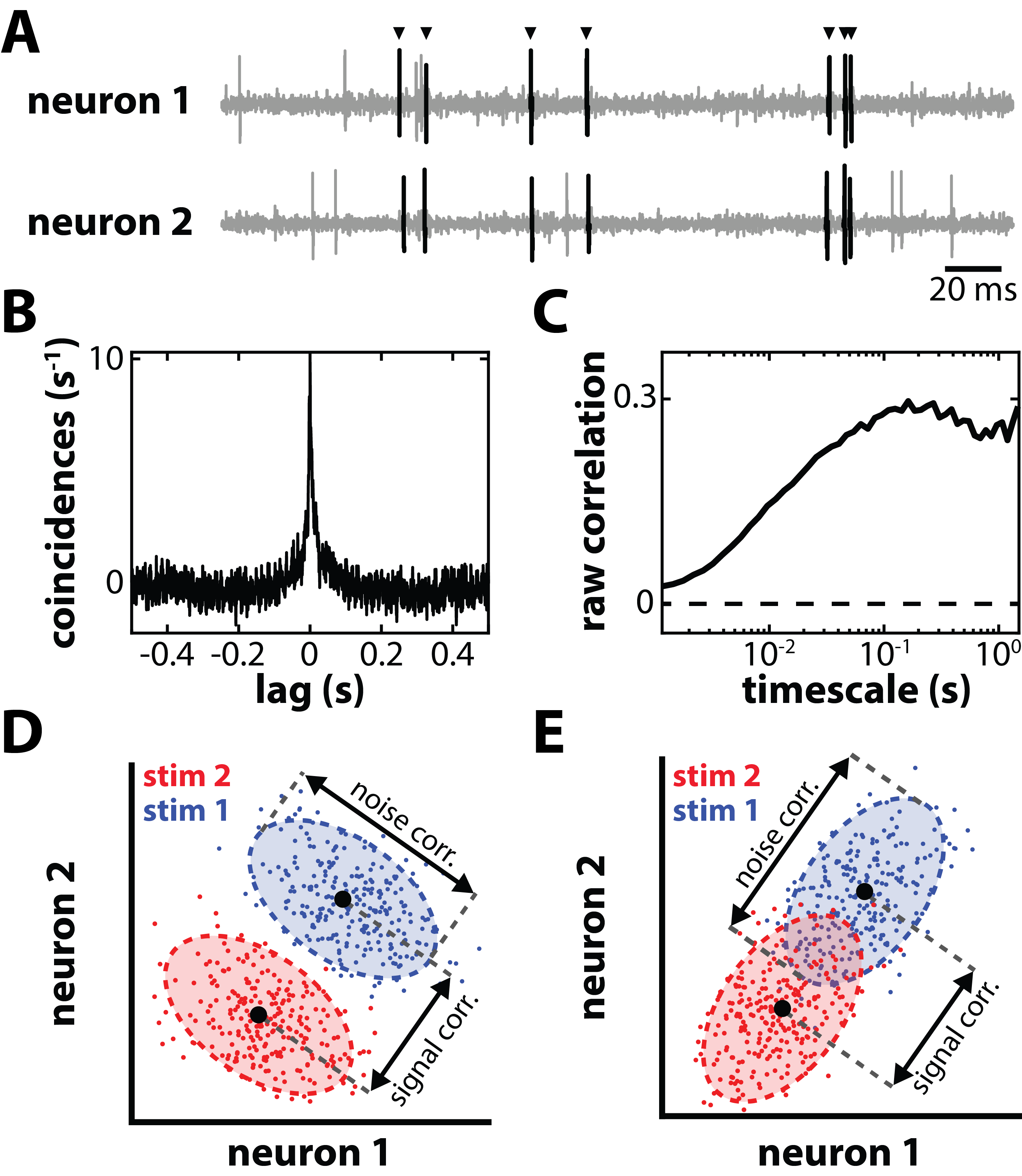
It is now widely agreed that perception and behavior are determined not by single neurons, but rather from the collective activities of large neural populations in the brain. Improvements in technology have allowed recordings from multiple neurons simultaneously, thereby revealing that neural activities are correlated (see panel A of figure to the right) and thus not independent of one another and is usually quantified by computing the cross-correlogram (CCG) between the activities of two neurons that were simultaneously recorded (panel B).
The CCG represents the number of coincident spikes above or below chance. Correlations can also be measured at multiple timescales ranging from a few (synchrony) to hundreds of milliseconds (co-variation of firing rates) (panel C). This is important because changes in correlated activity can be qualitatively different depending on timescale.
Moreover, correlations between neurons are highly plastic and have been shown to depend on multiple factors such as stimulus features and the internal state of the brain. We will henceforth refer to this as correlation plasticity. Correlations (raw) can be decomposed into two subtypes: signal and noise. Signal correlations quantify similarities between the mean responses of two neurons. In contrast, noise correlations quantify the similarity between the trial-to-trial variabilities of the neural responses to repeated presentations of a given stimulus.
We aim to understand how correlation plasticity affects population coding as well as the nature of the underlying mechanisms. Check out our recent publications on this topic.

Neuromodulation of sensory processing
There are important neuromodulatory pathways in sensory areas including: dopaminergic, neuradrenergic, cholinergic, and serotonergic. We are interested in how serotonergic pathways originating from the Raphe Nucleus can affect a sensory neuron's response to incoming stimuli. We use electrophysiological approaches in both brain slices and in whole animals to address the following questions:
- What are the cellular mechanisms by which serotonergic projections modulate excitability in sensory neurons.
- Do these mechanisms operate in whole animals?
- How will activation of serotonergic pathways affect responses to sensory input in whole animals.
Such an approach linking multiple levels will give a comprehensive framework for understanding the functional role of serotonergic modulation in vertebrates and the nature of the underlying mechanisms.

Functional role(s) of sensory adaptation
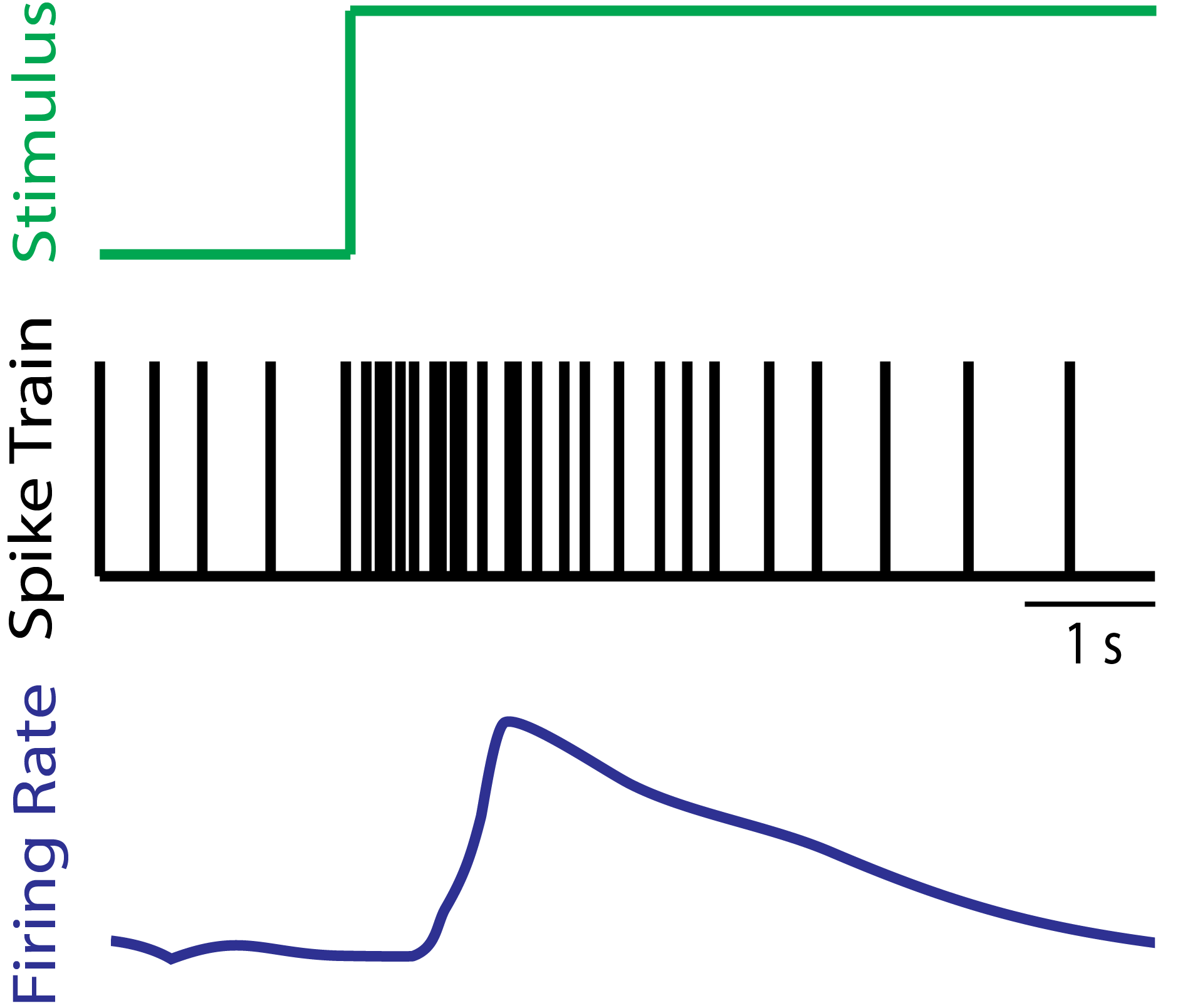
Our senses must efficiently process stimuli whose structure varies as a function of time and space. In order to to do, they continually adapt their response properties in order to optimize information processing. Examples of such adaptation abound (e.g., think of contrast adaptation) but the underlying mechanisms remain poorly understood to this day. We are interested in understanding how known mechanisms of adaptation can adaptively optimally encode stimuli with changing statistics.

Optimized coding of natural self-motion by vestibular pathways
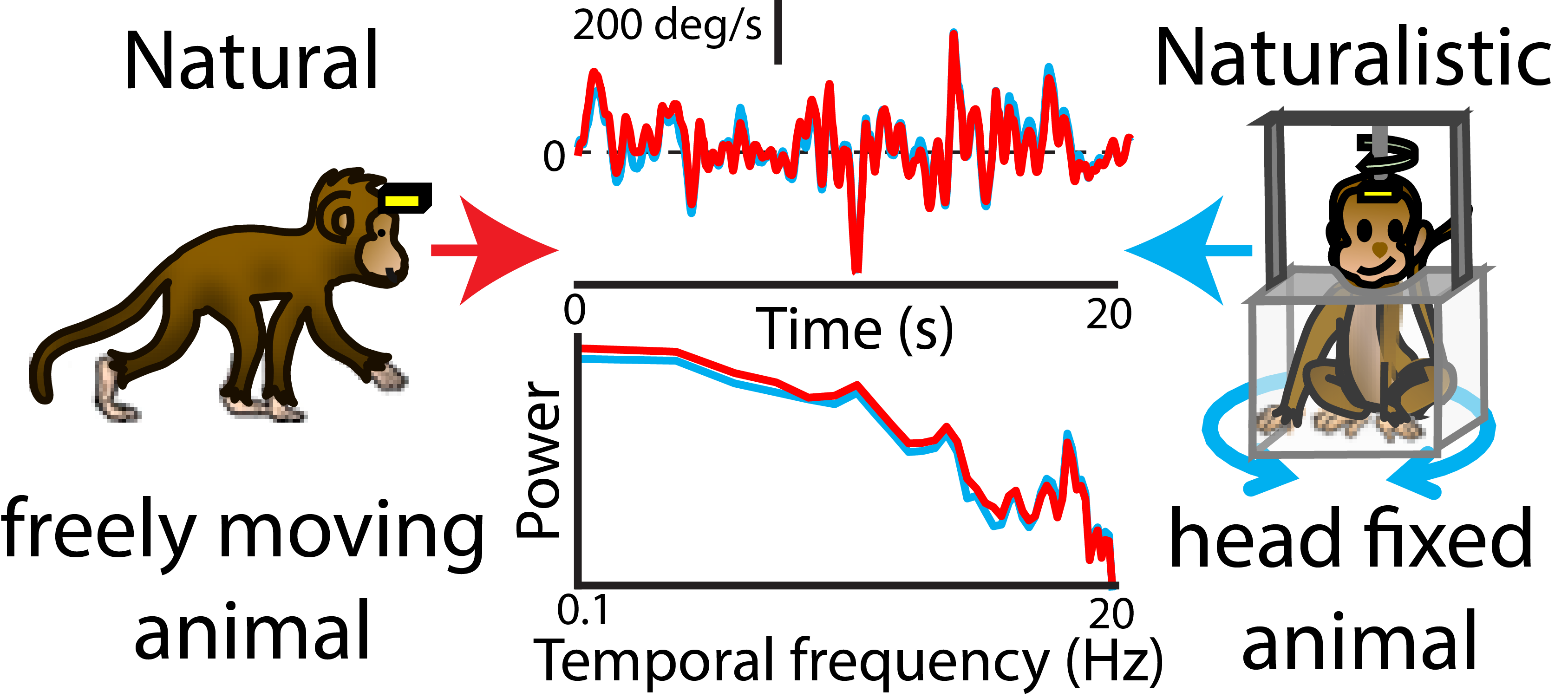
Growing evidence suggests that sensory pathways have adapted to the statistics of input found in the organism's natural environment in order to optimally encode them. Here we are interested in understanding whether this is also true for self-motion. To do so, we have first recorded the natural self-motion in freely moving NHPs and then used these to stimulate head-fixed animals (see Figure on the left). Our findings are that central vestibular neurons do encode such stimuli optimally through temporal whitening. Consult our recent paper on this topic. We are continuing this line of research by recording from downstream neurons within the Thalamus and cortex, as well as the abducens nucleus that controls eye movements, to understand how this information is decoded.
