Radiation Impact on Endodontic-Treated Teeth in Head-Neck Cancer Patients
E. Akbari, N. Karra, and F. Samim
Background: To date, there is uncertainty about whether head and neck cancer (HNC) patients with prior root canal treatments, who are clinically asymptomatic but have specific radiographic risk factors, should undergo additional treatment before receiving radiation therapy (RT). Considering the risk of oral complications ranging from mucosal to bone tissue impairments and weakened immune systems in RT patients, there is a lack of studies examining the impact of RT on asymptomatic root canal-treated teeth in HNC patients with specific defects.
Objective: This prospective observational cohort study in Montreal, Canada, seeks to describe the impact of RT on asymptomatic root canal-treated teeth in HNC patients with periapical radiolucency (PARL), underfilled and overfilled canals, widened periodontal ligaments (PDL), and defective coronal seals.
Methods: The records of 956 HNC patients who underwent RT between 2018 and 2022 were retrospectively reviewed. 286 patients with comprehensive dental, medical, and radiographic records were identified, of whom 122 had at least one root canal treatment. Demographic information, cancer diagnosis, treatment details, pre-RT dental records, radiographs, and all dental records during RT follow-up were thoroughly reviewed. 18 patients met the inclusion criteria. Updated radiographs (periapical and panoramic) and comprehensive dental examinations were used to document any symptoms of change in teeth with particular characteristics.
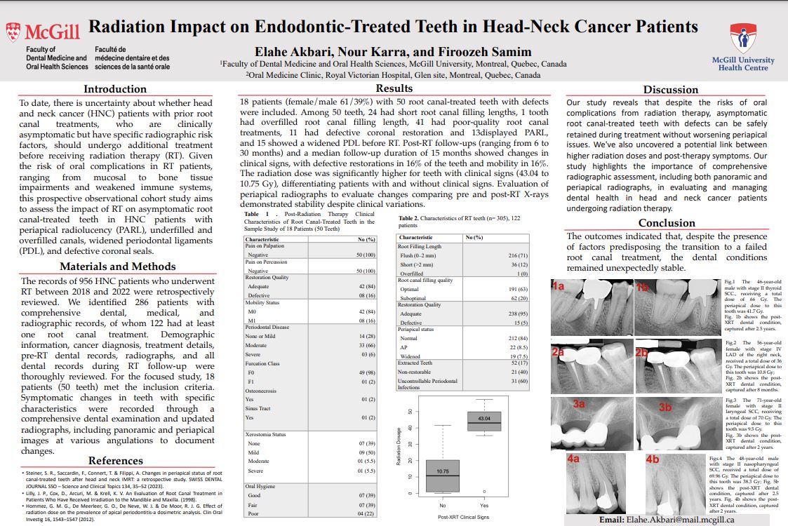
Results: 18 patients (female/male 61/39%) with 50 root canal-treated teeth with defects were included. Among 50 teeth, 24 had short root canal filling lengths, 1 tooth had overfilled root canal filling length, 41 had poor-quality root canal treatments, 11 had defective coronal restoration, 13 displayed PARL, and 15 showed widened PDL before RT. The median post-RT follow-up was 15 months. Clinical signs included defective restorations in 16% of teeth and mobility in 16% of teeth. The radiation dose was significantly higher for teeth with clinical signs (41.93 to 10.72 Gy). Evaluation of periapical radiographs to evaluate changes comparing pre- and post-RT X-rays demonstrated stability despite clinical variations.
Conclusion: The outcomes indicated that, despite factors predisposing the transition to a failed root canal treatment, the dental conditions remained unexpectedly stable.