

Enlarge Descriptive Card Log Book Entry (none)
Rodin Number: 52
E Number: (none)
Donor: Osler
Date: 1877
Size (H x W cm): 10.5 x 8
Six slices of lung show numerous worms within the airways (bronchi) (long arrow in magnified view A), causing distension and mucous plugging of their distal portion (short arrow).
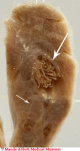

Click on caption to enlarge image.
Comment
Although the lungs are described as coming from a pig, Osler left no detailed record of pulmonary disease in such an animal. However, he did speak on the subject of lung worms in dogs at a meeting of the Montreal Veterinary Medical Association (of which he was then Vice President) on March 29, 1877. He had recently been asked to investigate an outbreak of disease in puppies at the Montreal Hunt Club. Eight of the affected animals which died underwent autopsy. A number of these showed irregularly sized, nodular swellings of the tracheobronchial mucosa, usually at or near an airway bifurcation. The swellings contained parasitic worms, which were not identified on the mucosal surface itself (an appearance identical to that seen in similarly affected dogs today, as can be seen in an image of the lower trachea (B), courtesy of the Ontario Veterinary College). Osler made a detailed description of the dog autopsies and a brief discussion of what was then known of nematode worm (Strongyloides) infection in animals.
Following correspondence with the editors of the London journal The Veterinarian (in which the material was subsequently published), Osler noted that the worm he observed was unlike those which had been described to that time and speculated that it was a new species. This was agreed upon by experts in the field and it was officially named Filaria (Filaroides) osleri by Cobbold in 1879. In 1929, a new genus was recognized and the name changed to Oslerus osleri, by which the organism is still known today.