NIKON CSU-X1 Spinning Disk Confocal
Applications: Intravital Imaging, 4D Confocal, Fast Kinetics, Widefield Fluorescence, Brightfield
Excitation:
- Four lasers lines for confocal imaging: 405nm, 488nm, 561nm, 640nm.
- Four LED lines for widefield fluorescence: 385nm, 488nm, 561nm, 621nm.
- Brightfield illumination.
Emission:
- Four single chanel emission filters: 455/50nm, 525/36nm, 605/52nm, 705/72nm.
- One quad chanels filter for high speed acquisition: DAPI/FITC/TRITC/CY5.
Detection:
- Confocal: Photometrics Prime BSI Express sCMOS Camera. 2048x2048, 95fps@11-bits/43fps@16-bits.
- Widefield and Brigthfield: Nikon Digital Sight 10, Color and Monochrome mode, 25 mm FOV, 23.9 megapixel.
Objectives: 10X/0.45 air, 20X/0.75 air, 16X/0.8 water dipping, 20X/1.0 water, 60X/1.42 oil.
Located in a biosafety level 2 room that include a BSC.
Image processing and analysis: Nikon imaging software (NIS) workstation and Bitplane Imaris 9.1.2
LEICA SP8 Laser Scanning Confocal and Multiphoton
Applications: 4D Confocal and Multiphoton Imaging, Spectral imaging, FLIM, FRET, FRAP modules, SHG
Excitation:
- Four solid state laser lines: 448 nm, 488 nm, 552 nm,
638 nm
- One tunable IR two-photons laser: 680-1080 nm
Detection:
- Two internal HyDs (high sensitivity detectors)
- Two internal PMTs
- One transmitted light PMT
- Two reflected light HyD
- One brightfield detector (Dodt gradient)
Objectives: 4x to 63x (including air, oil and multi-immersion)
Environmental control: CO2, temperature, humidity.
Image processing and analysis: Leica LAS X and Bitplane Imaris 9.1.2


PerkinElmer High Content Microscopes
Applications: Fully automated fluorescence and brightfield imaging of microplates (fixed or live samples) and standard slides. Automated high content image analysis.
Opera Phenix Plus automated microscope:
- From 6 well to 1536 well plates and standard slides.
- 5x, 10x, 20x air objective
 Perkin Elmer Opera Phenix Plus and Cell Explorer Station
 - 20X, 40x and 60X high numerical aperture water immersion objectives
- Brightfield, Digital Phase Contrast, Widefield or confocal (lens spinning disk)
- Five laser lines (375nm, 425nm, 488nm, 561nm, 640nm)
- Fourteen emission filters for wide or narrow emission window
- Four sCMOS 16 bit camera (2160px X 2160px)
- Fast frame-rate imaging capacity (up to 105 fps)
- Internal liquid handling capacity for fast kinetic acquisition (i.e., Calcium imaging)
- Connected to a plate handler, a liquid handler, a plate washer, an incubator and a bar scanner.
- Optional environmental control chamber (CO2, Temperature, Humidity)
Operetta automated microscope:
- From 6 well to 1536 well plates and standard slides
- 2x, 10x, 20x and 40x objectives
 Perkin Elmer Operetta automated microscope
 - A wide range of filter combinations
- Optional confocality (spinning disk)
- Optional environmental control chamber
- 300W xenon lamp
Browser-based image analysis software (Columbus)
Stereo-microscope (Leica M165)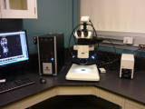
- Color camera 5Mp (Leica DFC450C)
- Epifluorescence (120W Mercury Halide lamp)
- Filter: green (GFP), red (RhodB), YFP, mCherry
- Reflected and Transmitted light set-up
- Leica LAS X acquisition software with multi focus and overlay module
Upright microscope (Leica DM1000)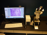
- Color camera 1.4Mp (Leica DFC310FX)
- Epifluorescence, Brightfield (120W Mercury Halide lamp)
- Filter: blue (A4), red (N2.1), green (L5)
- Leica LAS X acquisition software
Inverted microscope (Leica DMI 6000)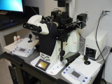
- Monochrome camera 2Mp (Leica DFC345FX)
- Epifluorescence, DIC, IMC (120W Mercury Halide lamp)
- Filter sets for DAPI, CFP, GFP and TxRed
- Leica LAS X acquisition software
- Eppendorf piezo system for microinjection/manipulation