Age/sex: 37-year-old female
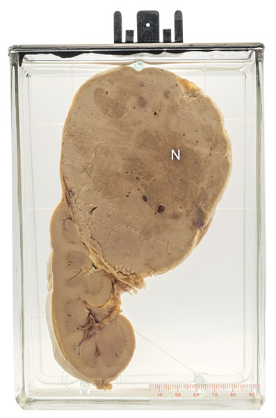
Size: 20.1 x 14.7 x 6.3 cm
The specimen shows a 10 cm well-circumscribed tumor in the upper pole of the kidney. Darker areas (N) likely indicate tissue death (necrosis).
Kidney carcinoma
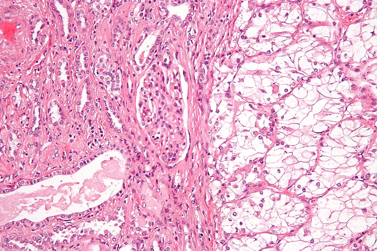
There are over 40 types of kidney carcinoma, each with a characteristic histologic appearance. The most common is called conventional adenocarcinoma. Often, it is composed of cells that contain abundant glycogen or fat. Both of these substances are dissolved during tissue processing for microscopic examination, leaving an empty space and giving rise to the alternate name clear cell carcinoma. The tumor is believed to arise from the proximal convoluted tubule of the renal cortex. Its cause is unclear; however, risk factors include cigarette smoking, obesity, and hypertension.
Most patients are older than 60 years. Many are asymptomatic, the tumor being discovered during abdominal imaging performed for another reason. Some patients present with flank pain, blood in the urine, or a palpable abdominal mass. Patients who have smaller tumors (< 4cm) that are confined to the kidney have a good prognosis following surgical resection (about 90% 5-year survival).
Below: Histologic appearance of clear cell carcinoma on the right; normal kidney tubules are on the left.
Source: Nephron. (2009). Clear cell renal cell carcinoma high magnification. https:// commons.wikimedia.org/w/index.php?curid=8255731