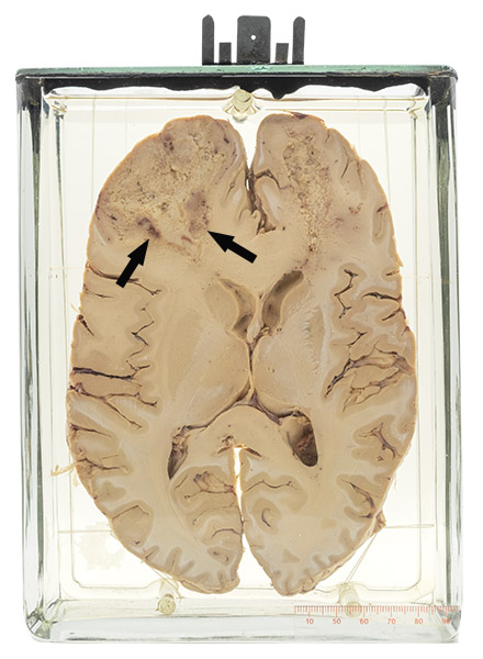
Age/sex: 63-year-old male
Size: 21.3 x 16.0 x 17.3 cm
A transverse slice of the brain shows a finely granular and focally hemorrhagic area in the white matter in the frontal lobe. Although this is most evident on the left side where the cancer originated (arrows), it is also present on the right where it spread.
Glioblastoma multiforme
Glioblastoma multiforme is the most common and most aggressive type of brain cancer. It is thought to arise from astrocytes (neuronal supportive cells) and usually originates in the white matter of a frontal lobe. Although the tumor can spread from its site of origin to the opposite lobe (as in this case), spread outside the brain (metastasis) is exceedingly rare. Most tumors develop de novo. However, some arise over a period of years from lower grade (relatively benign) tumors called astrocytomas.
Most tumors develop in individuals 50 – 70 years old and are manifested by headache, nausea/vomiting, seizures and/or personality change. Treatment includes surgery, chemotherapy and radiotherapy. However, usually these only delay progression of the cancer and death occurs in most people in 1 – 2 years.
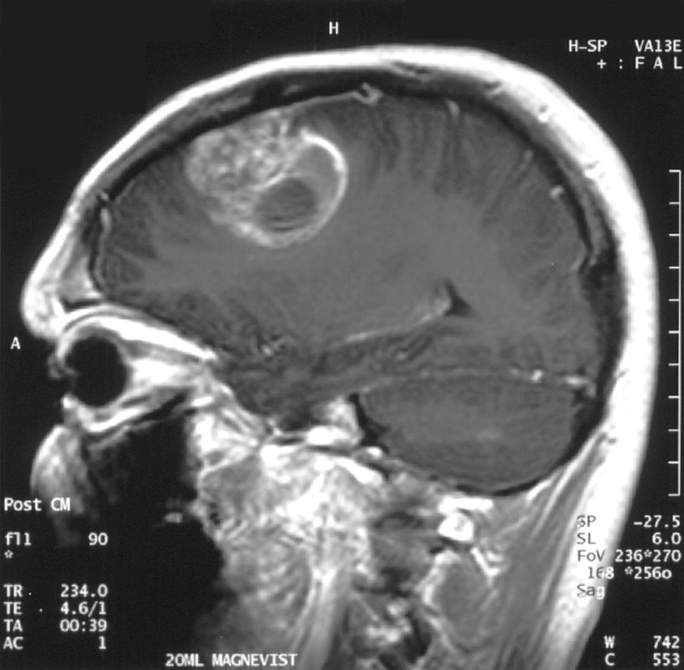
Below: MRI showing the typical appearance of a glioblastoma multiforme.
Source: Christaras, A. (2006). Glioblastoma - MR sagittal with contrast. Wikimedia Commons. https://commons.wikimedia.org/wiki/File:Glioblastoma_- _MR_sagittal_with_contrast.jpg