An aneurysm is the enlargement of an artery caused by a weakness in the arterial wall.
An aneurysm can develop in any section of the aorta. The location of the aneurysm determines its type:
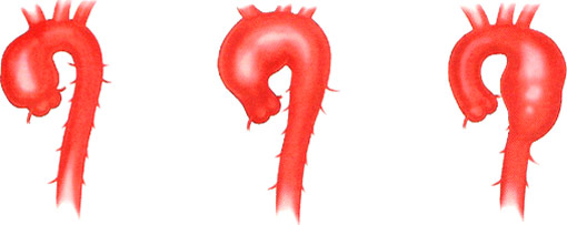
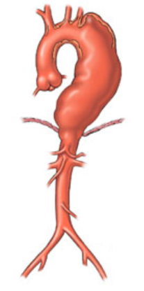
Thoracic Aorta Aneurysms occur in the portion of the aorta in the chest. A thoracic aortic aneurysm can develop in the aortic root, the ascending aorta, aortic arch or descending aorta.
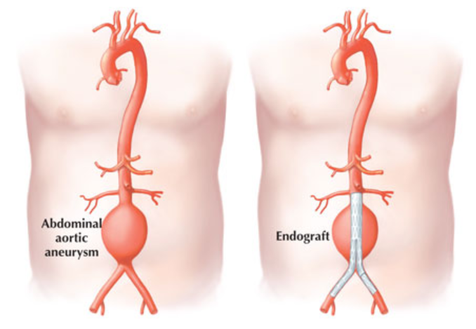
Abdominal Aortic Aneurysms occur in the section of the aorta that passes through the abdomen.
In some cases, an individual may have an abdominal aortic aneurysm and a thoracic aortic aneurysm.
Aneurysms can grow over time. As an aneurysm expands, it can start to cause symptoms. When an aneurysm gets too large, it can rupture (Acute Aortic dissection) and cause life-threatening bleeding or instant death — without any warning.
See the video: Aortic Aneurysm and Aortic Dissection
Who is affected by thoracic aortic aneurysms?
Men are more affected by aortic aneurysm than women, but women are more at risk of death because of the atypical presentation of the disease which often leads to a delay in getting diagnosed. It is difficult to evaluate the number of people who have an aneurysm, but we certainly know that the burden of disease increases with age.
How to diagnose:
Most aneurysms are often discovered during a routine health exam for some other unrelated condition. In other cases, an aneurysm is discovered when it has grown large enough to cause symptoms that send the person to the doctor. If an aortic aneurysm is suspected, your doctor may order the following tests:
- Chest x-ray: Often obtain for other indications, its usually the first imaging that may detect abnormalities of the aorta.
- Computed tomography (CT) scan: By using radiation, this test can detect the size and shape of an aneurysm.
- Magnetic resonance imaging (MRI): Alternative test to the CT scan for people who need frequent monitoring and to reduce their exposure to radiation.
- Echocardiography (an ultrasound of the heart): Using sound waves, this technique shows images of your heart and the ascending aorta. The doctor may also ask a transoesophageal echocardiogram to have a better view of your heart valve. Echocardiograms can also be used to screen family membesr suspected with aortic disease.
- Abdominal ultrasound: Helps to detect abdominal aneurysms and to follow up on the aneurysm. It is also an effective preventative screening tool for those individuals who are at high risk for aortic aneurysm.
- When the diagnosis of an aortic aneurysm is confirmed, your doctor may also propose Genetic screening if he is suspecting a connective tissue disease that could increase the risk of aortic dissection.
Treatments Goal:
The goal in treating aneurysms is to reduce the risk of rupture, which increases with aneurysmal size. However, if you have a small aneurysm, your doctor may propose treatment that will prevent your aneurysm from growing.
Your doctor may suggest:
- Medications: Aortic aneurysms are usually associated with high blood pressures that promote the weakness of the arterial wall. If you have high blood pressure, your doctor may initiate a Beta blocker (ex: Lopressor) or Angiotensin 2 receptor blocker (ex: Losartan) to lower your blood pressure. For people with Marfan's syndrome, it may slow the aortic dilatation rate. Your doctor will recommend maintaining a blood pressure lower than 140/90 or if you’re diabetic 130/80. Also, if you have an aortic abdominal aneurysm, a Statin will be introduced to your medication regimen. That medication will lower your cholesterol which can help reduce artery blockage and your risk of aneurysm complication.
- Monitoring with image: Your doctor will order regular imaging tests to evaluate the size of the aneurysm and determine whether the aneurysm is growing. You should expect an echocardiogram, CT scan or MRI, to be done between 3-6 months after you have been diagnosed with aortic aneurysm. The frequency and the choice of the tests will depend on the location, the size, and how fast the aneurysm is growing.
- Smoking cessation is recommended: As a known predisposing factor for abdominal aneurysm, your doctor may propose the use of a gum, nicotine patch or resources which could help you quit smoking.
- Screening for family members of patients with genetic aortopathy: Screening of the first-degree family members is indicated because the transmission rate is 50% in certain genetic disorder as Marfan. Also, genetic screening might help to clarify: (1) the nature of the disease; (2) the risk to the patient when the clinical diagnosis is uncertain. Your doctor may also suggest to do Imaging (Echocardiogram /MRI /CT SCAN) in family members suspected to have the disease.
- Avoid isometric exercise such as weight lifting, Valsalva maneuver (try to exhale when your airway is blocked) and heavy lifting. Those exercises promote high blood pressure, which can improve the aneurysm to grow.
Avoiding exercise involving a substantial isometric component, such as weightlifting, climbing steep inclines gymnastics, and pull‐ups.
Avoiding activities that are associated with rapid changes in atmospheric pressure (scuba diving, flying in unpressurized aircraft).
Performing aerobic activity at a moderate intensity (∼50% of aerobic capacity) and maintaining heart rate <100 bpm or <110 bpm for patients on and off β‐blockers, respectively.
For resistance training, higher repetitions at lower weights are recommended as opposed to lower repetitions with heavier weight, stopping before muscle fatigue.


Image by https://www.injurymap.com/free-human-anatomy-illustrations

- Promote a Healthy lifestyle: Aneurysms are linked to various cardiovascular diseases such as high cholesterol, hypertension and diabetic. Your doctor will recommend that you follow a low cholesterol and low salt intake diet, to drink with moderation, to be active, and maintain healthy weight. Link to Food Guide Canada
- Driving recommendation: The Canadian Medical Association suggest that patients with thoracic aortic disease to be precluded from private driving if the ascending aorta diameter is > 6.0 cm or the descending aorta diameter is > 6.5 cm, and restricted from commercial driving if the ascending thoracic aorta diameter is > 5.5 cm or the descending thoracic aorta is > 6.0 cm.
When an aneurysm reaches a critical size, approximately 5.5 cm, it is time for surgical treatment. However, depending of the rate at which your aneurysm is growing, your genetic tests result, your family history, the shape and size of your aneurysm, the location of your aneurysm, your symptoms or other medical conditions (ex: bicuspid valve, other aortic valve disease), you may be operated before the aneurysm reach the size of 5.5 cm.
Surgical Therapy:
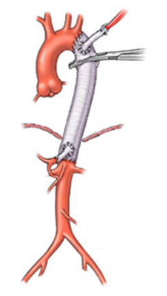
Open chest surgery

Depending on the location of the aortic aneurysm, the treatment could be open surgery or endovascular repair techniques. These are complex procedures that involve a team of doctors from different specialties working together.
During open surgical repair, an incision is made in the chest or abdomen, depending on the location of the aneurysm. During the procedure, the bulging, diseased area of the aorta is lined with a synthetic graft that is stitched in place to connect it with the normal aorta on either side of the defective area. When the procedure is completed, the new, synthetic section of the blood vessel functions like a normal, healthy aorta.
The procedure generally takes between 3 and 5 hours, and the hospital stay averages between 5 to 10 days. Some patients stay at a rehabilitation facility for a short time to complete their recovery. Most people return to their normal activities in 6 to 12 weeks. Open surgical repair is a proven treatment with very good long-term results.
The type of surgery that you will receive will depend on the location of the aneurysm. When you meet your surgeon he will explain the risk surrounding the surgery and the structure of your aorta or valve that will be affected by the surgery.
Endovascular Repair
The endovascular repair is the preferred treatment for abdominal aortic aneurysm if you are a candidate for this approach. Instead of an open chest surgery, the surgeon makes a small incision in the groin area to access the arteries that connect to the aorta. During the procedure, the surgeon will be using catheters that will deliver a stent graft through the blood vessel to the site of aneurysm. Once it is securely in place, the stent-graft creates a new passageway for blood flow without pushing on the aneurysm. Over time, the aneurysm will shrink because of the lack of pressure on it. Typically, the procedure takes 1 to 3 hours and you can go home in a few days after the procedure. Recovery time is generally faster with this intervention. Most people return to their normal activities in 2 to 6 weeks after the procedure. After endovascular procedure, you will need regular follow-up imaging scan to monitor the graft position.
Image of endovascular repair: