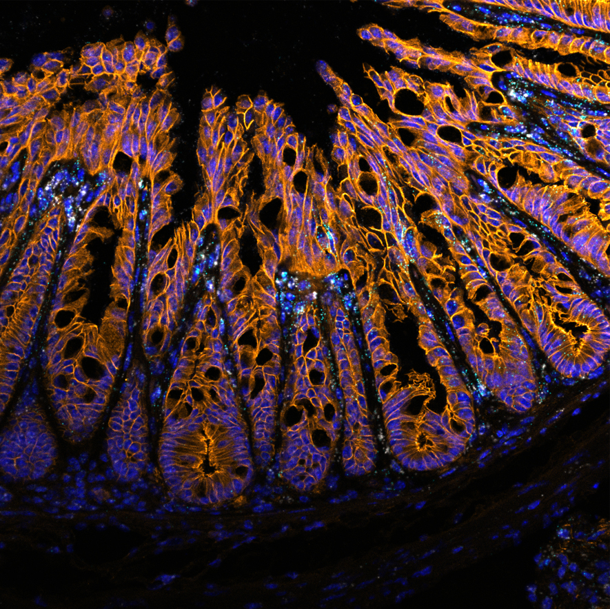
1st place Jessica Pei
Confocal image showing fluorescent in situ hybridization (FISH) stain in mouse colon. Cd109 (White) and Ifi44 (Cyan) mark an inflammatory fibroblast population expressed at the tops of colonic crypts in mice that were infected with Citrobacter rodentium. The intestinal epithelial cells are labelled with E-cadherin (Orange) and cell nuclei are labelled with DAPI (Blue).
Acknowledgements: Dr. Samantha Gruenheid, Dr. Alex Gregorieff, MUHC Molecular imaging platform
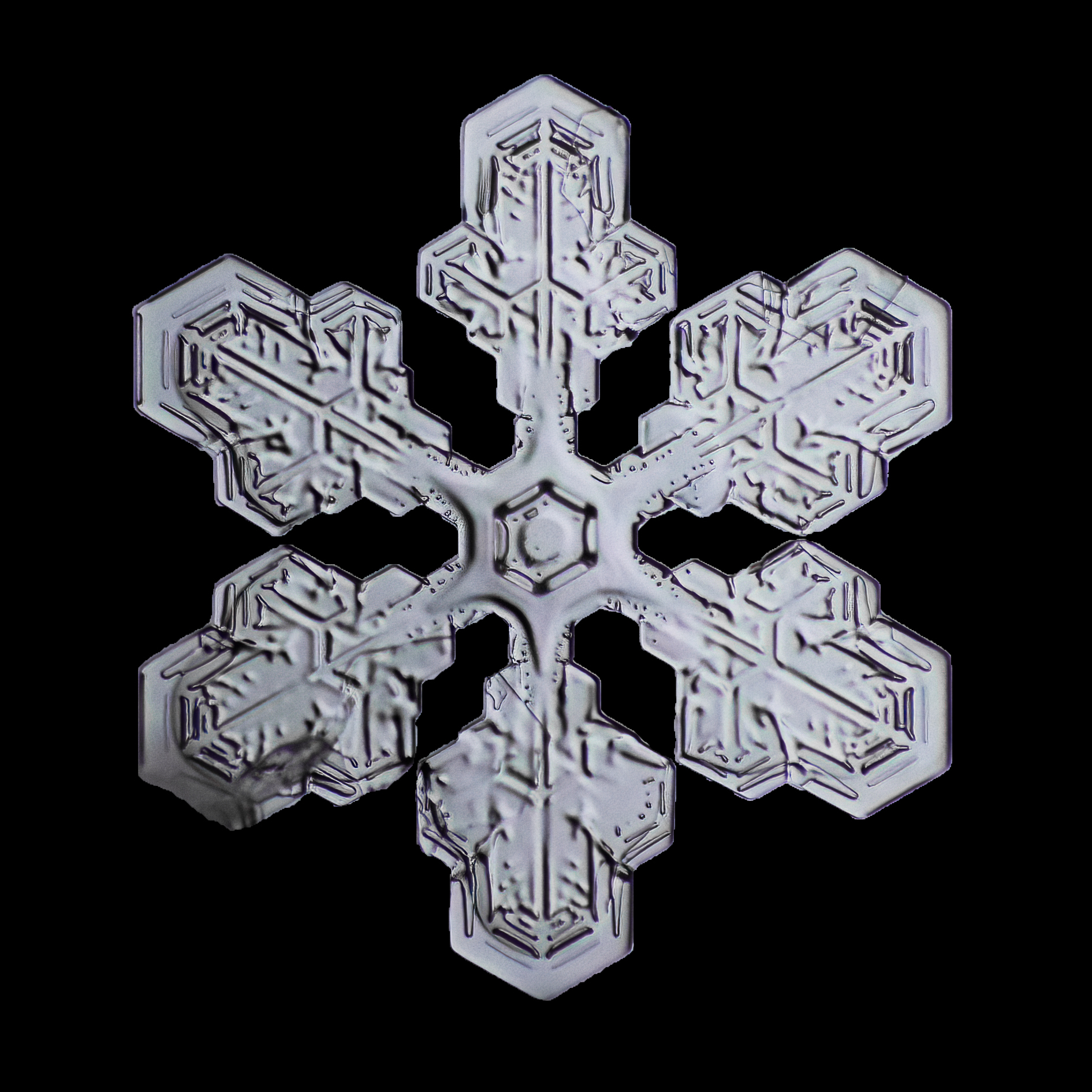
2nd place - Marcus Saldanha
This is a Stellar Dendrite Snowflake, one of the many kinds that fall to the ground when it snows.
The capturing process began by using a Canon t3i attached to an inverted Canon EF 28-135mm lens (this is where the microscopic effect comes from). Since there is a narrow depth of field, I took ~40 images then stitched them together in photoshop. Once rendered, I spent a few hours in lightroom adjusting the colours and a few minor details to get the final version.
Next time it snows, I ask you to take a closer look at the little pieces of art falling on your jacket and just maybe you will be as intrigued as I was when I started macrophotography. If you want to see more snowflake photos, check out my instagram @saldanhaphotography.
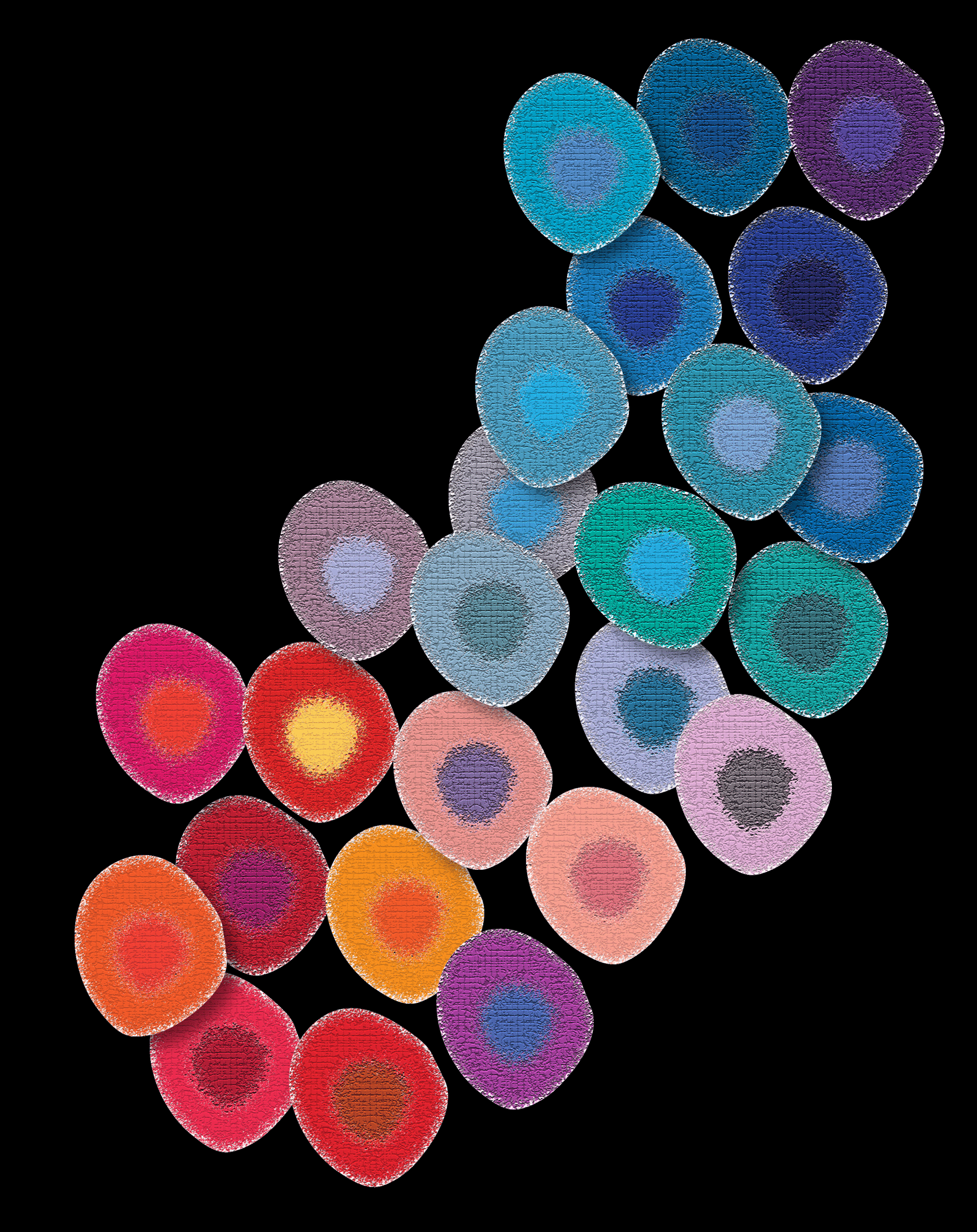
3rd place Dr. Judith Mandl and Dakota Rogers
Naïve CD4+ T cells are heterogeneous with respect to their strength of reactivity to self (coded from red to purple). We show that this self-reactivity spectrum is established by pre-wired transcriptional and chromatin states that are partially imprinted during thymic development.
Submissions

 .
. 


 .
. 