Abbott Specimen 50
Specimen Card Nomenclature
Stenosis of right ventricle at lower bulbar orifice. Conus a separate chamber with infective endocarditis of margins of orifice, wall of conus and pulmonary valve and large infective thrombus in conus cavity
International Classification of Diseases
Congenital right ventricular outflow tract obstruction
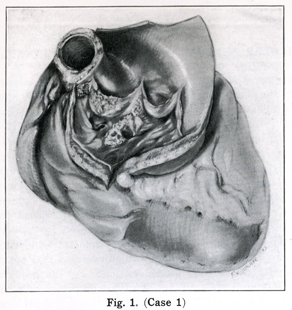
Atlas Illustration
Plate XVII, Figure 1
Donor
Dr. Thalheimer
Date
1928
Age
Middle aged woman
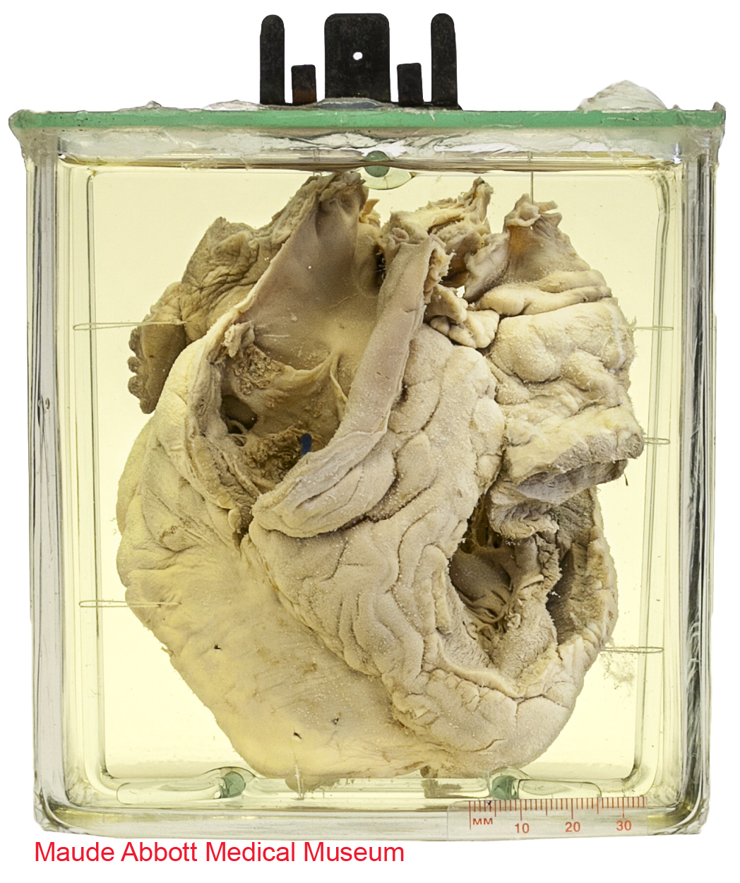
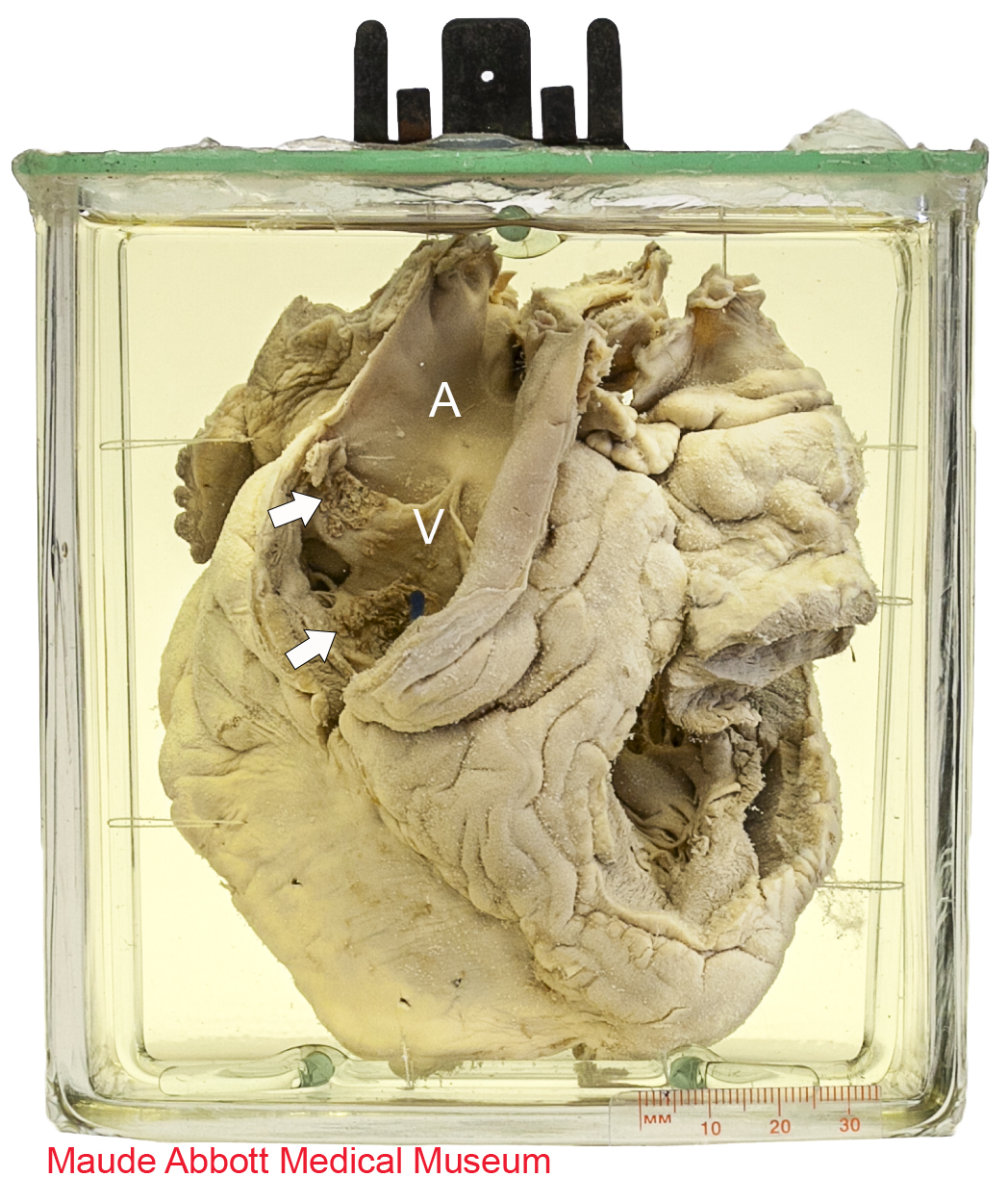
Description
Front view shows a structurally normal pulmonary artery (A) and valve (V). A blue rod is present in a markedly narrowed right ventricular outflow tract below the valve. Small granularities (arrows) can be seen on its upper surface and on one of the pulmonary valve leaflets, consistent with small thrombotic vegetations of infectious endocarditis.
Comment
Pulmonary blood flow can be compromised by stenosis below, at or above the pulmonary valve. It may be accompanied by a septal defect, such as a patent foramen ovale, allowing blood to shunt from the right to the left side of the heart. As with any cardiac anomaly that causes an alteration in normal blood flow, this increases the risk of infection, as occurred in this case.
Atlas Illustration
Plate XVII, Figure 1