
Abbott Specimen 74
Specimen Card Nomenclature
Patent ductus arteriosus with infective pulmonary endarteritis and embolic abscesses of the lungs
International Classification of Diseases
Patent arterial duct with infectious endarteritis
Atlas Illustration
Plate XIII Figure 4b
Donor
Drs. Hamilton & Grunner
Date
1913
Age
Nineteen years
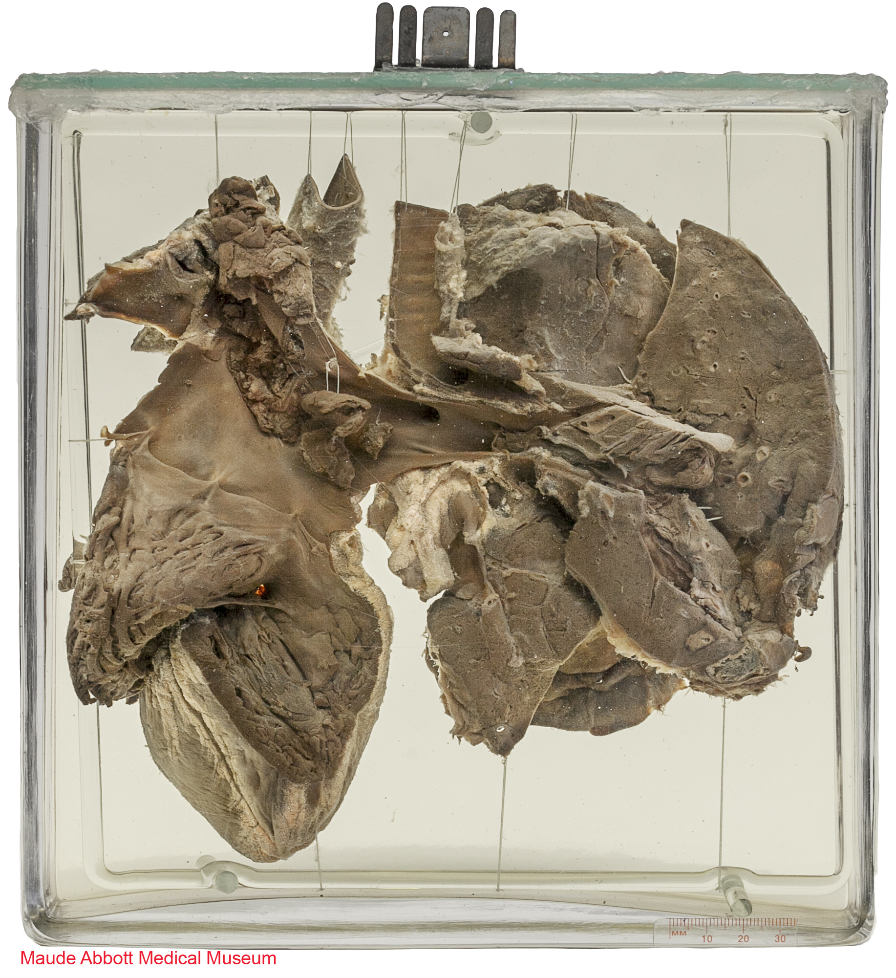
Description
The specimen shows an opened right ventricle (V). The pulmonary artery above is partly occluded by thrombus (T) which extends into the aorta through a previously patent ductus arteriosus (arrow). The right lung has been sliced to show several 0.5 - 1.0 cm abscesses (arrows), representing embolic spread of infection from the pulmonary artery.
Comment
The sixth aortic arch contributes the proximal portions of both pulmonary arteries. On the right side, the distal part loses its connection with the aorta and regresses; on the left, it persists as the ductus arteriosus. The latter provides an intrauterine conduit for blood to flow from the right heart to the aorta, bypassing the non-functional lungs.
The ductus maintains its patency in fetal life by the action of prostaglandins produced by the placenta and the ductus itself. After birth, these mediators decrease precipitously. Combined with an increase in blood PO2, this usually results in closure of the ductus functionally within 2-3 days and anatomically within 2-3 weeks (forming the fibrous ligamentum arteriosum). Failure of the ductus to close (patent ductus arteriosus) can result in a left to right shunt (higher pressure aortic blood flowing into the pulmonary artery) and eventually pulmonary hypertension.
The anomaly occurs in about 8 of 1000 premature babies and 2 of 1000 full term ones. the functional and clinical significance depend on the size of the ductus lumen, larger ones being more serious. One particularly serious complication, as in this case, is infection of the ductal endocardium (endocarditis).

![]() Enlarge View
Enlarge View
Atlas Illustration
Plate XIII Figure 4b

Figure 12:30. Langman, Jan. Medical Embryology: Human Development⎼Normal and Abnormal. Baltimore: The Williams & Wilkins Company, 2nd ed, 1969.
