Abbott Specimen 52

Specimen Card Nomenclature
Aortic atresia
International Classification of Diseases
Aortic atresia
Atlas Illustration
None
Donor
Dr. Gruner (Montreal Maternity Hospital)
Date
1913
Age
Fifteen days
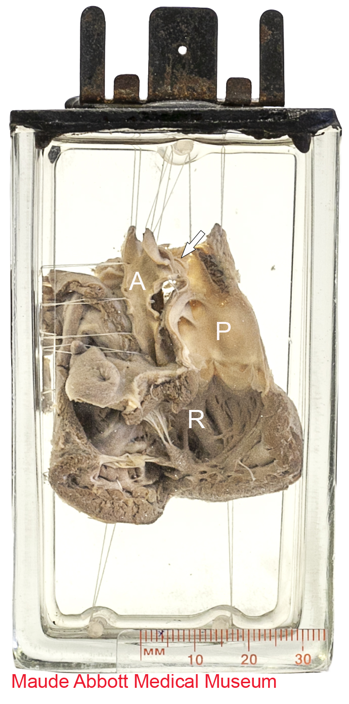
Description
Front view shows a dilated right ventricle (R) and a pulmonary artery (P) that is about four times the size of the aorta (A). A small patent ductus arteriosus (short arrow) connects the two.
Comment
Aortic valvular atresia is part of a spectrum of obstructing malformations of the left ventricular outflow tract, which can occur in sub-valvular, valvular, or supra-valvular regions. When the valve is the site, it can be monocuspid, bicuspid, tricuspid (often dome shaped) or quadricuspid. It can be an isolated anomaly or associated with a hypoplastic left ventricle.