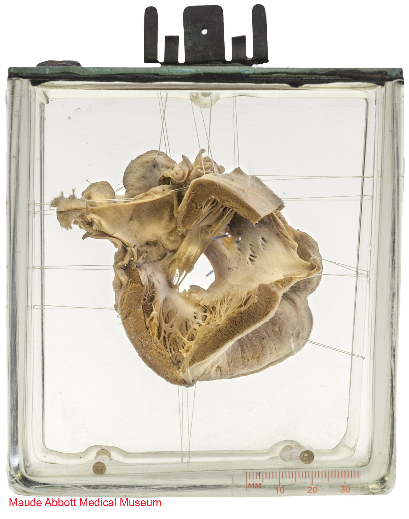
Abbott Specimen 75

Specimen Card Nomenclature
Common auriculo-ventricular orifice with persistent o.i. and cleavage of mitral and tricuspid segments over defective ventricular septum. Imperfect double mitral orifice
International Classification of Diseases
Atrioventricular septal defect
Atlas Illustration
None
Donor
Dr. R. M. Taylor
Date
1914
Age
Infant
Description
Front view shows a 1.3 cm defect in the atrioventricular septum. A small atrial septal defect is also indicated by a blue rod.
Comment
The interventricular septum has a complex development involving the ventricular muscle, endocardial cushions and bulbus cordis. The inferior (muscular) portion develops by growth of myocardium laterally on both sides of the developing heart with simultaneous resorption (trabeculation) on the inner portion. The central portion of this trabeculated mass fuses to become the muscular interventricular septum.
The septum thus formed is incomplete superiorly, leaving an interventricular defect which closes by growth and fusion of tissue derived from the endocardial cushions and the conal ridges. This forms the membranous portion of the interventricular septum. Abnormal growth of the endocardial cushions/conal ridges results in the development of a common atrioventricular valve and a communication that spans the atrial and ventricular components of the atrioventricular septum, as seen in the specimen illustrated here. Approximately half of the infants born with Down syndrome (which this child had) have significant congenital heart disease; the most common abnormality is an atrioventricular septal defect.
Figure 12:16. Langman, Jan. Medical Embryology: Human Development⎼Normal and Abnormal. Baltimore: The Williams & Wilkins Company, 2nd ed, 1969.

Figure 12:18. Langman, Jan. Medical Embryology: Human Development⎼Normal and Abnormal. Baltimore: The Williams & Wilkins Company, 2nd ed, 1969.
(A) six weeks (12 mm); (B) beginning 7th week (14.5 mm); (C) end of 7th week (20 mm)
