
Enlarge
Donor: Dr. Rhea
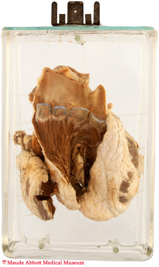
Date: 1922
Size (H x W x D cm): 18 x 12 x 6
The opened right ventricle shows four distinct pulmonary valve leaflets.
History: not known.
Comment: The semilunar (pulmonary and aortic) valves develop as swellings of the endocardial cushions on the inner aspect of the truncus arteriosus. When the four cushions fuse in the midline to divide the ventricular chambers, two additional swellings appear centrally. Continued growth is accompanied by excavation on the truncal side, resulting in the formation of sinuses between the presumptive leaflets and aorta/pulmonary artery wall. The precise mechanisms by which additional leaflets are formed or leaflets fail to develop are unclear. However, some cases of bicuspid aortic valve have a genetic basis (autosomal dominant with incomplete penetrance).
Supernumerary leaflets (typically a quadricuspid pulmonary valve as in this case) occur rarely (0.001 to 0.003 live births) and are usually unassociated with symptoms or complications.