
Enlarge
Donor: Unknown
Date: 1951
Size (H x W x D cm): 13 x 15 x 5
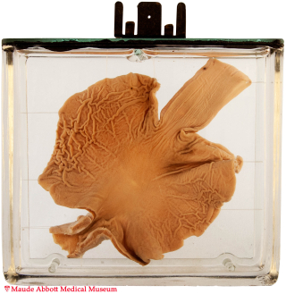
Moderately severe muscle thickening can be seen in the pyloric region (A, arrows). The operative site was excised for sectioning and is not evident.

A. Click on image to enlarge.
History: Ten week-old boy born 3 weeks before term. Although reported to have “taken his feeding poorly from birth”, development was said to be normal. He died one month after undergoing Ramstedt’s operation for pyloric stenosis. It is not clear if the stenosis or the operation to correct it were factors in this, since the infant also had polycystic kidneys, which may have caused renal failure.
Comment: Congenital pyloric stenosis results from hypertrophy of the pyloric muscle in utero. The cause is unclear and appears to have both genetic and environmental influences. The enlarged muscle impairs gastric emptying leading to vomiting with dehydration in the short term and “failure to thrive” (weight loss) more chronically.
The abnormality is easily treated by myotomy (cutting the hypertrophic muscle), a procedure pioneered by the German surgeon Ramstedt in 1911.