
Enlarge
Donor: Royal Victoria Hospital
Date: 1951
Size (H x W x D cm): 12 x 9 x 3
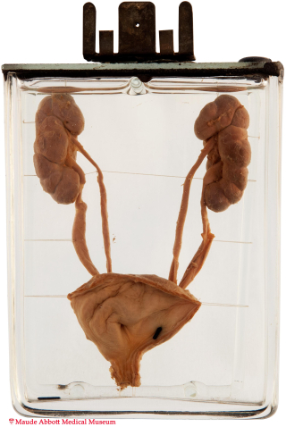
The kidneys show fetal lobulation, but are otherwise unremarkable. Two renal pelvices can be seen on each side (more clearly on the left, arrows in A). Each gives rise to a ureter, which join near the bladder wall (back view, B).


A. Click on image to enlarge. B. Click on image to enlarge.
History: Still born (unknown gestational date). Concomitant anencephaly and spina bifida.
Comment: Ureteral development begins around the 4th week of gestation as a ureteric bud arising from the mesonephric (Wolffian) duct near the cloaca. It is believed that ureteral duplication occurs if this bud either splits or arises twice. Frequently, the associated kidney is divided into upper and lower lobes, which blend imperceptibly into each other. Occasionally (as in this case), the division gives rise to two separate pelvices in the same kidney; rarely, there are two separate kidneys.
The condition is not uncommon, affecting about 1% of the population. In most cases, the two ureters join to form a single ureter, which enters the bladder normally (partial or incomplete double ureter); in this instance, there are usually no associated complications or symptoms. Rarely, the two ureters are completely separate, each draining into the bladder, or one into that structure and the other into the vagina or urethra. Individuals so affected may have incontinence or hydroureter/hydronephrosis, sometimes complicated by infection.