
Enlarge
Donor: Unknown
Date: Unknown
Size (H x W x D cm): 18 x 12 x 6
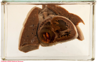
The right superior pulmonary vein (A, short arrow) courses upward to join a dilated superior vena cava (A, long arrow). The right atrium (A) is markedly dilated.

A. Click image to enlarge.
History: Unknown
Comment: The pulmonary veins develop as an evagination of the dorsal wall of the atrium. This grows toward the developing lung buds, which are initially drained by the splanchnic plexus into the cardinal veins. Ultimately, the splanchnic connections regress leaving four atrial derived veins (right and left, upper and lower lobes).
Anomalous pulmonary venous drainage occurs when pulmonary venous blood flows directly into the right heart or a systemic vein. It may be partial or total. Many drainage patterns have been described; they can be grouped into suparcardiac, cardiac and infracardiac types. Examples of partial flow include drainage from the left upper lobe to the left brachiocephalic vein and from the right lung to the inferior vena cava (in which case the right lung is commonly hypoplastic).
Total anomalous drainage is less common than the partial form and is necessarily associated with an atrial septal defect or patent ductus arteriosus. Frequently, the veins join behind the heart and form a somewhat dilated sac which courses to the vena cava, right heart or infradiaphragmatic vessel (see Specimen 18). Stenosis of the abnormal vein (either intrinsic or by compression) can obstruct blood flow and lead to right heart failure.