
Enlarge
Donor: Royal Victoria Hospital
Date: 1926
Size (H x W x D cm): 23 x 20 x 10
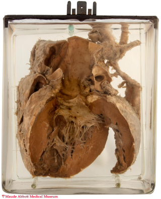
The descending aorta is narrowed to a thin cord 0.5 cm in diameter and 4 cm in length (A, arrows) beginning just beyond the origin of the left subclavian artery. The ascending aorta is dilated (8 cm diameter) and the left ventricular wall measures 2 cm in thickness, both features consistent with increased pressure. The aortic valve is bicuspid (a finding present in about 50% of cases) and mildly fibrotic.

A. Click on image to enlarge.
History: Fourteen year-old boy. Detailed history unknown, but said to have had a “cerebral death”, possibly related to hemorrhage secondary to hypertension.
Comment: Aortic coarctation occurs in about 1 to 3 per 10,000 births. The stenosis may be located proximal or distal to the insertion of the ductus arteriosus (Fig. 12-33). In the pre-ductal form, blood flows to the lower body via a persistent patent ductus. Because other cardiovascular abnormalities are frequently present, this form usually becomes evident at birth or in early infancy.
In the post-ductal form (as in this case), blood flows to the lower body by an enlarged collateral circulation originating in the proximal aorta and coursing through the mammary and intercostal arteries (not seen in this preparation). Depending on the severity of the stenosis, symptoms may not become evident until adulthood.