
Enlarge
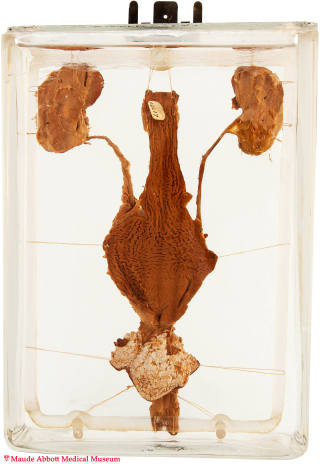
Donor: Jewish General Hospital, Montreal
Date: 1940
Size (H x W x D cm): 20 x 14 x 6
A posterior view shows a dilated rectum with a 1 mm depression (A, arrow) near its junction with the anal squamous mucosa. Its communication with the urethra is seen on the front view (B, arrow).

A. Click image to enlarge. B. Click image to enlarge.
History: Thirty-eight day-old infant boy; history unknown.
Comment: In early development, the terminal portion of the hindgut abuts the cloaca, from which it is separated by a cloacal membrane. Formation and downward growth of the urorectal septum divides the cloaca into an anterior portion (the primitive urogenital sinus) and a posterior anorectal canal. At about 7 weeks, the urorectal septum reaches the cloacal membrane to form the primitive perineum. The cloacal membrane is then known as the urogenital membrane anteriorly and the anal membrane posteriorly. At the end of the 9th week, the anal membrane is situated between the rectum and an ectodermal depression (the proctodeum, which forms the lower part of the anal canal). Subsequent rupture of the membrane leads to communication of the anal canal with the rectum. Failure of this rupture leads to imperforate anus, often accompanied by rectal fistula with the vagina, urinary bladder, or urethra.