
Enlarge
Donor: Royal Victoria Hospital
Date: 1930
Size (H x W x D cm): 21 x 7 x 4
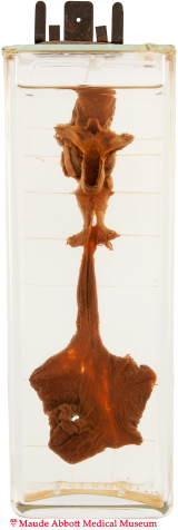
The proximal esophagus is seen as a 2 x 1 cm blind ending pouch. The red rod demonstrates a fistula between the distal esophagus (front view) and the trachea (back view, A).

A. Click on image to enlarge.
History: Five day-old infant boy; history and additional anomalies unknown.
Comment: see specimen 39.