
Enlarge
Donor: Dr Chase
Date: 1932
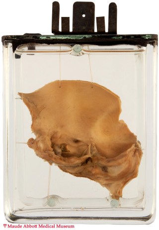
Size (H x W x D cm): 11 x 9 x 3
Abbott No 76
The aortic valve consists of two leaflets, each of which shows mild to moderate fibrosis. The aorta itself appears normal, despite distal coarctation (not shown in this preparation).
History: Thirty-four year-old primipara who had signs of heart failure during pregnancy. She underwent Caesarian section at 8 months followed by the delivery of a healthy baby. However, she (the mother) was found unexpectedly dead in bed six nights later.
Comment: See Specimen 08.
This specimen is part of a larger one (now lost) illustrated in Maude Abbott’s Atlas of Congenital Cardiac Disease (Plate VIII, Fig. 2). In this sketch, one can see the heart with a hypertrophied left ventricle as well as the aortic coarctation.
Plate VIII, Fig. 2. Atlas of Congenital Cardiac Disease. Drawing by H Blackstock, Medical Art Department, McGill University.
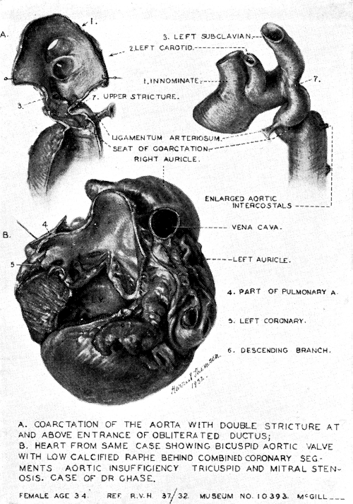
Plate VIII Figure 2.