
Enlarge
Donor: Royal Victoria Hospital
Date: 1916
Size (H x W x D cm): 25 x 13 x 11
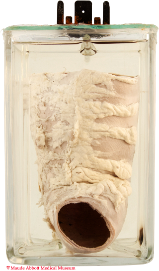
A segment of the transverse colon is markedly dilated (9 x 6 cm on top view, A), but otherwise structurally unremarkable.
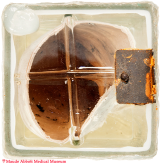
A. Enlarged of a segment of the transverse colon.
History: Four year-old boy; portion of sigmoid colon removed surgically.
Comment: The nervous system begins as a plate like sheet of thickened ectoderm in the mid-dorsal region of the embryo (neural plate). Laterally, it develops neural folds, which become elevated and fuse to form a midline neural tube. Neural crest cells at the apex of the tube migrate into various body tissues and differentiate into sensory ganglia of the spinal and cranial nerves, as well as contributing to a variety of other important structures such as the cardiac endocardial cushions and pharyngeal pouches.
During the 12th week of intrauterine life, cells from the neural crest also migrate into the presumptive colon to form Auerbachs and Meissner’s plexuses. In Hirschsprung disease, this migration is incomplete or absent. The part of the colon which lacks these nerve networks cannot relax, causing physiologic obstruction and colon dilatation. In most individuals, the disorder affects only the distal colon; in about 5%, the entire colon is affected.
Although the condition was first described in the late 1700s, it is named after Harald Hirschsprung, a Danish physician who described two infants who died of the disorder in 1888.