

Enlarge
Donor: Alexandra Hospital, Point St Charles, Quebec
Date: 1935
Size (H x W x D cm): 14 x 10 x 5
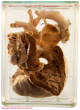
A 1.3 cm defect is evident just below the aortic valve. A 1 cm patent foramen ovale can be seen to the right. The left ventricle is markedly dilated. A view from behind (B) shows right ventricular dilatation and hypertrophy (wall thickness - 0.5 cm), findings consistent with a left to right shunt. The ligamentum arteriosum (arrow) is prominent but closed (the connection with the pulmonary artery lumen showing no orifice).
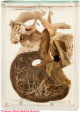
A. Click on image to enlarge. B. Click on image to enlarge.
History: Two and 1/2 year-old boy. History unknown.
Comment: The interventricular septum has a complex development involving the ventricular muscle, endocardial cushions and bulbus cordis. The inferior (muscular) portion develops by growth of myocardium laterally on both sides of the developing heart with simultaneous resorption (trabeculation) on the inner portion. The central portion of this trabeculated mass fuses to become the muscular interventricular septum. The septum thus formed is incomplete superiorly, leaving an interventricular defect which closes by growth and fusion of tissue derived from the endocardial cushions and the conal ridges. This forms the membranous portion of the interventricular septum. Abnormal growth of the endocardial cushions/conal ridges results in the most common form of ventricular septal defect, as seen in the specimen illustrated here.
The abnormality is often isolated, but may be associated with other cardiac anomalies, as in this case. It is frequently present in Down syndrome. Small defects are not uncommon; the vast majority of these close spontaneously during infancy as the heart grows. Larger defects may lead to significant left to right shunt and the development of pulmonary hypertension. In long standing cases, the shunt may reversed (right to left), in which case the addition of unoxygenated venous blood in the left ventricle is associated with decreased systemic PO2 and cyanosis. The turbulent flow across the defect is a risk factor for the development of infection (endocarditis).