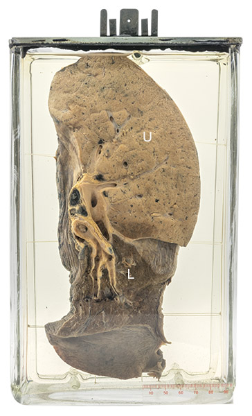
Age/sex: 57-year-old male
Size: 24.2 x 15.2 x 7.1 cm
The specimen shows a slice of right lung whose lower lobe (L) is somewhat collapsed, but otherwise normal. The upper lobe (U) has a tan color due to complete consolidation (filling) of airspaces by white blood cells.
Lobar pneumonia
This form of pneumonia is caused most often by the bacterium Streptococcus pneumoniae. Inhalation of the organism is followed by an outpouring of edema fluid and inflammatory cells (neutrophils) from alveolar capillaries into the adjacent alveolar airspaces, resulting in an area of more or less uniform consolidation. When extensive – sometimes affecting an entire lung lobe - this can be associated with spread of the bacterium into the blood (sepsis) and death. The consolidation results in a dull instead of a resonant sound when the chest wall overlying the area of pneumonia is percussed.
Leopold Auenbrugger (1722 – 1809) is credited with popularizing the use of percussion―tapping the chest or abdomen and listening to the ensuing sounds―in the diagnosis of disease. According to tradition, he learned its value as a boy by having to find the level of wine in his father’s casks. In 1761, he published his observations on the medical use of the procedure in a book titled New Invention by Means of Percussing the Human Chest as a Sign of Detecting Obscure Diseases in the Interior of the Chest.
“… with the finding of the resonance impaired or totally suppressed in the affected thorax, the patients are more rarely afflicted with a cough. … with active movements, suffocation attacks them … respiration and speech are now interrupted … the pulse becomes rapid and irregular … these are the signs which indicate a hardening of the lung.” (Auenbrugger’s description of clinical findings in lobar pneumonia.)
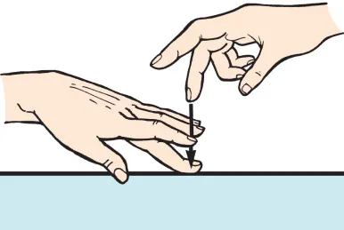
Below: An illustration of the percussion technique used by Auenbrugger.
Source: Fig 1.5. Method of chest percussion. From “The History and Physical Examination.” (2019), Thoracic Key. https://thoracickey.com/the-history-and-physical-examination-4/