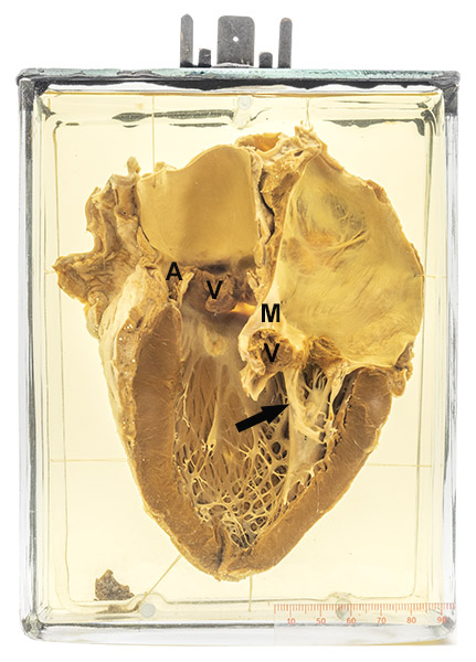
Age/sex: 37-year-old female
Size: 19.3 x 15.4 x 6.7 cm
The specimen has been opened to show the left ventricle with mitral (M) and aortic (A) valves. The chordae tendineae of the former are thickened and fused (short arrow) consistent with chronic rheumatic valve disease. Large brown vegetations (V) are present on the surface of both valves; some of these appear to be associated with destruction of the underlying valve leaflet.
Infectious endocarditis
Infectious endocarditis is defined as inflammation of the lining of the inner surface of the heart caused by a microorganism (usually a bacterium). The mitral and the aortic valves are the most common sites. The valves can be normal (in which case the causative bacterium is usually virulent, such as Staphylococcus aureus) or fibrotic (scarred). The latter are most often the result of rheumatic fever affecting the mitral valve or of degenerative disease (“aging”) involving the aortic valve.
Toxins secreted by the bacteria and enzymes released from the body’s neutrophils that accumulate to counter the infection damage the underlying valve and can lead to perforation or sometimes its complete destruction. Thrombus (blood clot) develops on the injured area forming a “vegetation”. Because of the physical “clapping” of valve leaflets during routine beating of the heart, fragments of these vegetations can become detached and spread to various organs where they impact within small arteries and cause tissue death (infarcts).
William Osler, one of McGill’s best-known physicians, described the clinical manifestations and corresponding pathologic findings of endocarditis in a series of lectures in England in 1885. Some of the hearts from which he learned about the disease can be seen in the Museum’s Osler cabinet.
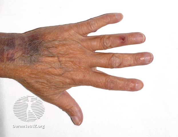
Below: An Osler node on the fourth finger, a classic finding of endocarditis caused by embolization of a fragment of thrombus (vegetation) that has formed on a valve leaflet.
Source: Osler nodes. (2006). DermNet. https://dermnetnz.org/topics/osler-nodes-and-janeway-lesions