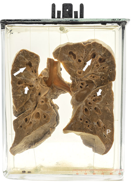
Age/sex: 3½-year-old male
Size: 19.3 x 14.5 x 6.5 cm
The specimen shows the right and left lungs with saccular (long arrows) and cylindrical (short arrows) bronchiectasis in all lobes (see Specimen 14). Although such disease is often more severe in the upper lobes in cystic fibrosis, in this specimen it is fairly uniform from apex to base. The small, irregularly shaped pale areas in the lung parenchyma (P) represent foci of bronchiolitis and pneumonia.
Cystic fibrosis
Cystic fibrosis is a genetic disorder that affects multiple organs, particularly the lungs, pancreas, and intestines. It is caused by a mutation in the gene whose product is a transmembrane protein that regulates chloride ion transport across cell membranes. Abnormalities of this protein lead to changes in various organ secretions, such as mucus in the lung and digestive secretions in the pancreas. These in turn lead to pathological findings of pulmonary bronchiectasis and pancreatic atrophy with corresponding clinical manifestations of cough (caused by retained “sticky” mucus) and weight loss (due to the inability to digest food).
Although the disease had been recognized since the 1500s, it was not known by the name cystic fibrosis until 1936 following studies by Dr. Dorothy Andersen (1901 – 1963). Although she had received a medical degree from Johns Hopkins in 1923 and subsequently trained as an intern in surgery at Strong Memorial Hospital in Rochester NY, she was not granted a residency position because of her sex. Instead, she undertook a career of research (interestingly partly in congenital cardiac disease, similar to that of Maude Abbott).
Following an autopsy on a child thought clinically to have celiac disease, Andersen found pancreatic cysts and fibrosis; subsequent investigations led her to the conclusion that the combination of these abnormalities and lung cysts (which we now know to be bronchiectasis) formed an entity which she termed cystic fibrosis.
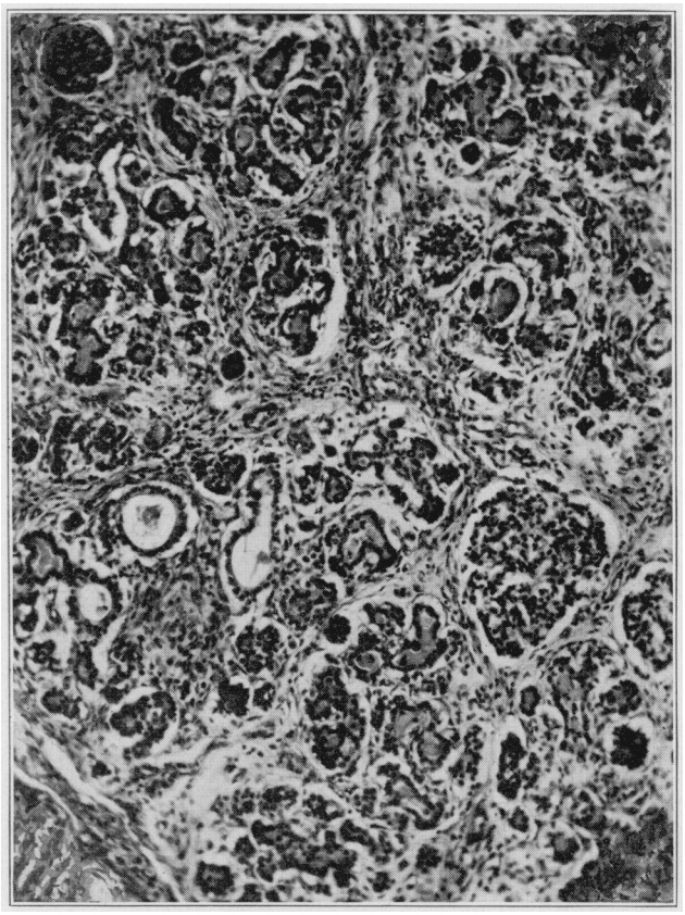
Below: Photomicrograph of the pancreas in a patient with cystic fibrosis who died at the age of 8 weeks. This image was published in Dr. Dorothy Andersen’s original paper describing multiple cases of what she termed “cystic fibrosis”.
Source: Andersen DH. Cystic fibrosis of the pancreas and its relation to celiac disease: A clinical and pathological study. Am J Dis Child. 1938;56(2):344–399. doi:10.1001/archpedi.1938.01980140114013