
Age/sex: 66-year-old female
Size: 18.4 x 12.5 x 5.1 cm
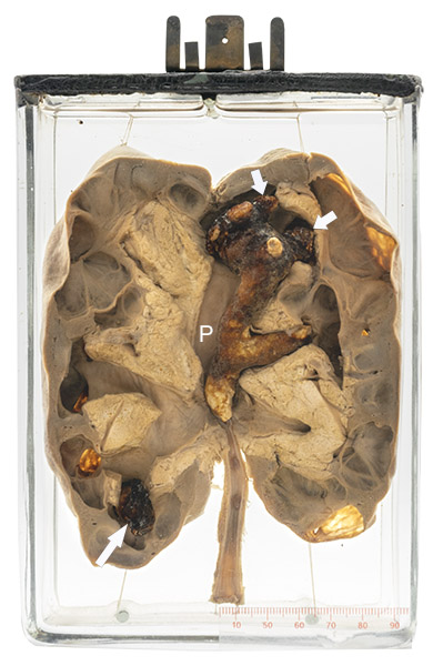
The kidney has been cut in its mid-portion and separated in two halves. A branching, somewhat nodular brown structure can be seen filling the upper pole calyces (short arrows) and pelvis (P). A smaller structure can be seen in the lower pole (long arrow). Both of these would be quite hard to the touch.
Kidney stones
After the gallbladder, the kidney is the most common organ for the formation of stones (calculi). They can be composed of different substances, with different causes and clinical associations. The most common is calcium, usually in combination with oxalate or phosphate. It is typically associated with a high blood calcium level such as seen in hyperparathyroidism. Other substances are urate (associated with gout) and struvite (associated with urinary tract infections).
Individual stones range in size from less than 1 mm to over 10 cm. Larger ones tend to form a cast of the renal pelvis/calyx (the region of the kidney that collects urine before it is drained via the ureter to the bladder). Because of the normal shape of this region, the stones are often branched, as in specimen displayed; hence the descriptor “staghorn”.
Stones can stay in the kidney for years without causing pathological or clinically evident disease. In many cases, however, they interfere with urine flow and cause scarring of the adjacent kidney (as in the specimen displayed). They also predispose to infection. Small stones or fragments of larger ones can pass from the pelvis down the ureter to the bladder, sometimes causing severe pain (renal colic).
Below: A staghorn calculus made of struvite.
Source: Gaillard, F. (2010). Staghorn calculus. Radiopaedia. https://radiopaedia.org/cases/staghorn-calculus-photo
