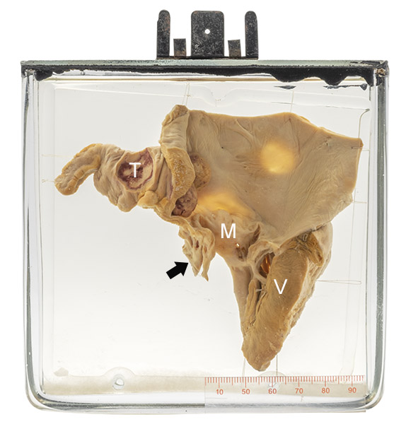
Age/sex: 40-year-old female
Size: 13.3 x 13.7 x 3.0 cm
The specimen consists of the left atrium with a portion of the left ventricle (V) underneath. The mitral valve (M) is moderately thickened. The chordae tendineae (arrow) that attach it to the myocardium are fibrotic and fused together. The atrial appendage is filled with thrombus (T).
Mitral stenosis and atrial thrombosis
The pathologic findings are those of rheumatic mitral stenosis (see Specimen 43). The increased atrial pressure caused by hindrance of blood flow across the stenotic valve is associated with slower and abnormal blood flow in the atrium. Both of these processes predispose to thrombus formation. Atrial fibrillation - an irregular and frequently fast heart rhythm sometimes seen with mitral stenosis - is also a factor in some cases.
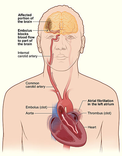
The thrombus rarely causes problems within the appendage. However, fragments of it may be dislodged and travel via the left ventricle and aorta to an artery elsewhere in the body (embolization) where they can occlude the vessel and cause tissue death (infarction). The most common organs so affected are the kidneys, spleen, and brain. Nowadays, treatment with blood thinners (anticoagulants) greatly decreases the risk of atrial appendage thrombosis and complicating embolization.
Below: The mechanism by which atrial thrombus can cause stroke through embolization.
Source: National Heart Lung and Blood Institute. (2013). Atrial fib stroke. Wikimedia Commons. https://commons.wikimedia.org/wiki/File:Atrial_fib_stroke.jpg