
Age/sex: 77-year-old female
Size: 14.0 x 14.0 x 6.5 cm
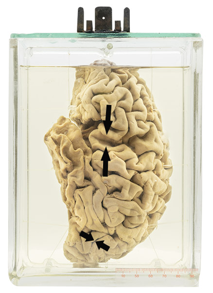
Left brain hemisphere with the frontal lobe at the top. Its sulci are increased in width (long arrows), indicating loss of adjacent cortical neuronal tissue (compare with normal sulci (short arrows) in the occipital lobe at the bottom of the specimen).
Pick’s disease
Pick’s disease (frontotemporal dementia) is an uncommon condition that presents with language, thinking and personality/behaviour changes. It is caused by the accumulation of tau proteins in neural cells. These proteins are normally responsible for maintaining the structural stability of neural axons. In Pick’s disease, their abnormal accumulation leads to cell damage and, eventually, death. When many neuronal cells have died, the cortical gyri are thinned and the adjacent sulci widened , such as in the specimen shown. Disease particularly affects the frontal lobes.
In many people, the disease is associated with inheritance of an abnormal gene. It tends to be seen at a younger age than Alzheimer’s disease (sometimes in patients as young as 20 – 30 years) and usually progresses to death within 3 to 5 years.
Arnold Pick (1851 – 1924) was a Czech psychiatrist who described the clinical and corresponding pathological findings in 1892.
Below: A neuron shows accumulation of amorphous, lightly staining purple material (tau protein) next to its nucleus (arrow).
Source: Degeneration of the frontotemporal lobes of the brain. Swiss Medica. https:// www.startstemcells.com/frontotemporal-dementia.html
![]()