Age/sex: 27-year-old female
Size: 20.5 x 11.9 x 7.7 cm
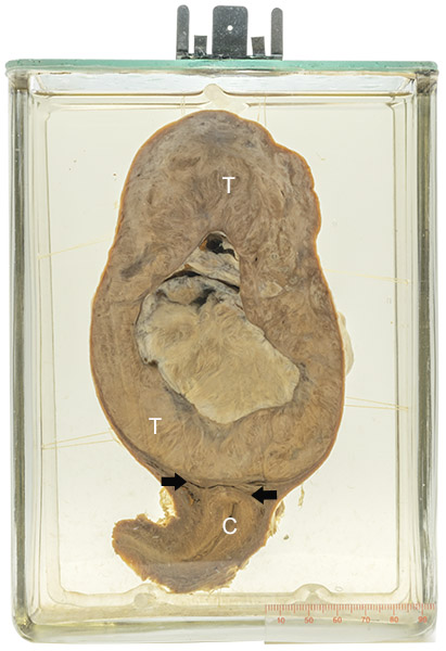
The uterus has been opened to show the endocervical canal (C) and a uterine cavity (arrows) that is markedly compressed by a 12 x 8 cm tumor (T) above. The center of the tumor is partly cystic and white, indicating that it is dead (necrotic). The patient was known to have had the tumor for five years before it was excised.
Leiomyoma
Leiomyomas are benign neoplasms of smooth muscle. Because they are often quite firm to the touch, they are also known as fibroids. Their most frequent location is the uterus, in which they are very common: it has been estimated that they develop in as many as 40 % of Caucasian women and 80% of African-American women.
The tumors range in size from microscopic to over 20 cm. Although they may be solitary (as in the specimen shown), multiple tumors are frequent. They can be seen at all ages and are not uncommon in young women. Because of changes in blood estrogen and progesterone levels, they may increase in size during pregnancy and decrease in size after menopause.
Most leiomyomas cause no symptoms. However, large ones can cause menstrual irregularities and are rarely associated with infertility. Sometimes they cause urinary frequency (wanting to urinate often) or constipation, due to pressure on the bladder or colon. Occasionally, a tumor outgrows its blood supply and dies (necrosis), in which case there may be acute pelvic pain.
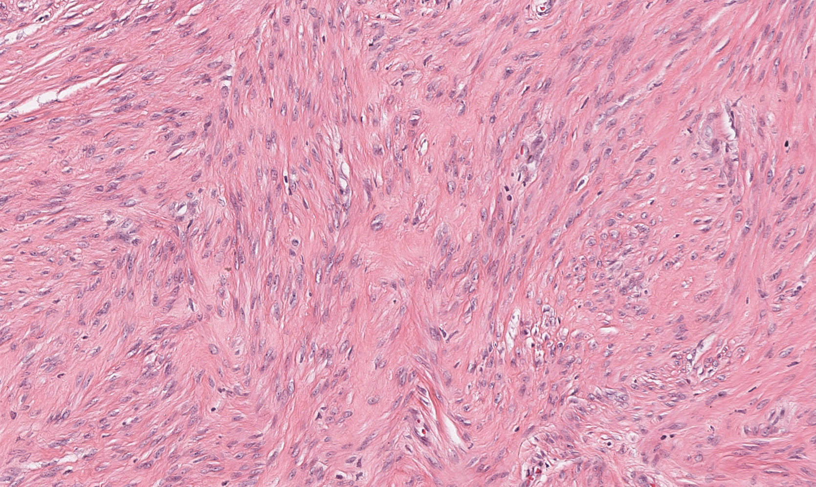
Below: Histologic appearance of a leiomyoma showing intersecting fascicles of cells that have elongated (spindle-shaped) nuclei.
Source: Rajaram, A. (2023). Leiomyoma.