
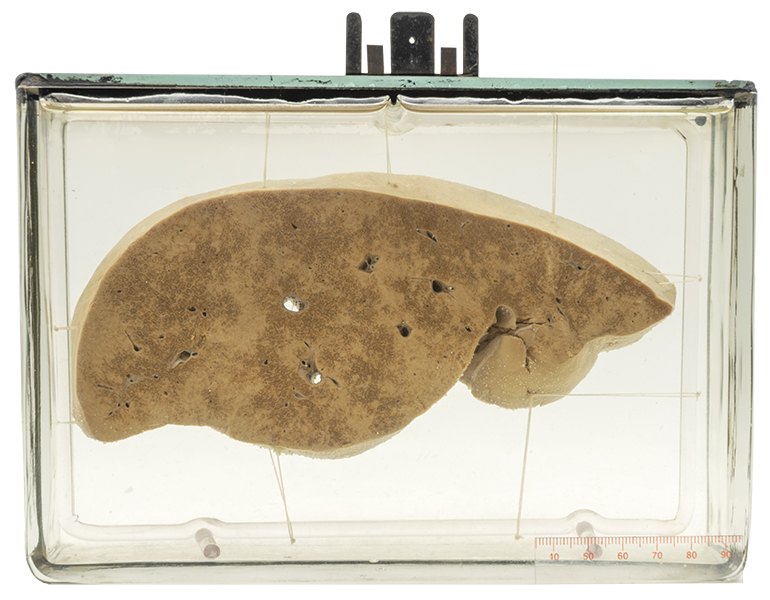
Age/sex: 45-year-old male
Size: 14.8 x 20.6 x 7.2 cm
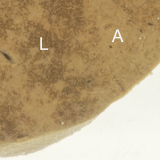
The specimen consists of a slice of liver that has a variable appearance, some areas tan in color and others paler. The pale regions are deposits of amyloid. In some areas (best seen on a magnified view), there is minimal deposition (L); in others, the liver appears to be completely replaced (A) by amyloid.
Amyloidosis
Amyloidosis is characterized by the accumulation of an abnormal protein in extracellular tissue. This can be localized to a single organ or can involve many sites throughout the body. The abnormal protein is derived from a normal body protein that becomes misfolded into a structure called a ß-pleated sheet. The misfolding is associated with an inability of body cells to metabolize and dispose of the protein. With continued accumulation, the abnormal protein (called amyloid) compresses adjacent cells and leads to their death. If enough cells are affected, organ function is affected and clinical signs and symptoms develop, such as liver failure in the patient whose specimen is shown.
Although all amyloid looks the same histologically and has the same underlying ß-pleated sheet structure, it can be derived from several normal body proteins. The underlying diseases and clinical findings associated with these precursor proteins are different. Two of the most common precursors are:
- immunoglobin light chain protein (AL amyloid), associated with plasma cell myeloma (a form of lymphoma (see Specimen 2);
- serum amyloid A protein (AA amyloid), produced by liver cells in chronic infectious or inflammatory diseases such as tuberculosis and rheumatoid arthritis.
Below: Histologic appearance of hepatic amyloidosis; L - liver cells; A - amyloid.
Source: Nephron. (2011). Hepatic amyloidosis. Wikimedia Commons. https:// commons.wikimedia.org/wiki/File:Hepatic_amyloidosis_-_intermed_mag.jpg
