
Age/sex: 20-year-old female
Size: 19.1 x 8.0 x 4.1 cm
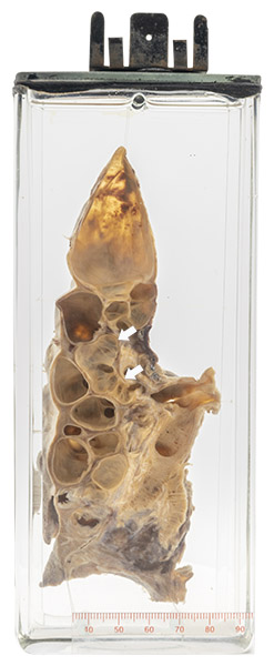
The specimen consists of a lower lung lobe in which the parenchyma has been completely replaced by multiple cystic structures of variable size and shape, each representing part of a dilated bronchus. Some (arrows) are elongated and have thin transverse folds on their walls, indicating that they are bronchi rather than a parenchymal abnormality such as a healed abscess.
Bronchiectasis
Bronchiectasis can be defined as permanent dilatation of a bronchus. It may be slight over its whole length (cylindrical), more prominent and sinuous in shape (varicose), or large and localized (saccular).
There are many causes (including cystic fibrosis, such as seen in Specimen 13). The specimen shown here was most likely the result of a childhood infection such as pertussis (whooping cough). It is thought that such infection damages and weakens the airway wall, which leads to an increased risk of further airway infection and, eventually, dilatation.
Rene Theophile Hyacinthe Laënnec (1781–1826) was the first physician to describe bronchiectasis in detail. Although he was also the first to report a number of other diseases, he is best known for his 1819 book on clinical applications of the stethoscope: De l’auscultation médiate ou Traité du Diagnostic des Maladies des Poumon et du Coeur:
“I recalled a well-known acoustic phenomenon: if you place your ear against one end of a wood beam the scratch of a pin at the other end is distinctly audible. It occurred to me that this physical property might serve a useful purpose in the case I was dealing with. I then tightly rolled a sheet of paper, one end of which I placed over the precordium (chest) and my ear to the other. I was surprised and elated to be able to hear the beating of her heart with far greater clearness than I ever had with direct application of my ear. I immediately saw that this might become an indispensable method for studying, not only the beating of the heart, but all movements able of producing sound in the chest cavity.”
Below: An illustration of Laënnec’s stethoscope: 1) instrument assembled; 2) and 3) two portions of the instrument in longitudinal section; 4) detachable chest piece; 5) earpiece unscrewed; 6) transverse section.
Source: Roguin A. Rene Theophile Hyacinthe Laënnec (1781-1826): the man behind the stethoscope. Clin Med Res. 2006 Sep;4(3):230-5. doi: 10.3121/cmr.4.3.230.
