
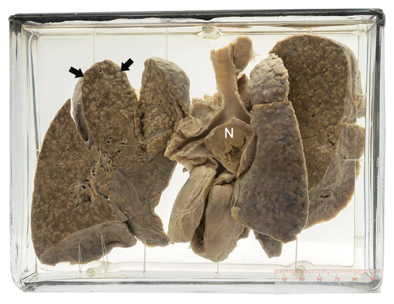
Age/sex: 3-year-old female
Size: 16.2 x 21.7 x 7.6 cm
The lungs of this child show numerous small white nodules in all lobes (arrows). One large focus of necrotic tissue beneath the trachea (N) represents the presence of infection in a lymph node.
Miliary tuberculosis
This form of tuberculosis develops when Mycobacterium tuberculosis gains access to the blood and spreads throughout the body, causing innumerable foci of infection, often in many organs. These foci range in size from the minute (visible only with the microscope) to about 2-3 mm in diameter, as in this case. They do not usually become larger because when they reach this size the amount of disease is such that most patients die.
John Jacob Manget (1652 - 1742) described this form of disseminated tuberculosis in 1700, likening the size and appearance of the foci of disease to millet seeds (from the Latin miliarius - related to the millet seed).
Below: Chest CT of a middle-aged woman with miliary tuberculosis. Notice the innumerable, tiny, grain-like foci throughout both lungs.
Source: Mugdal, P. (2015). Miliary tuberculosis.
