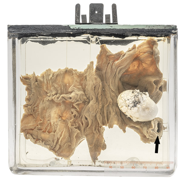
Age/sex: 41-year-old female
Size: 13.5 x 14.5 x 8.4 cm
A segment of small bowel contains a white, egg-shaped gall stone. The proximal bowel lumen is markedly dilated (compare with the unopened distal bowel lumen — arrow).
Gallstone ileus
Gallstone ileus is a rare complication of cholelithiasis (stones in the gallbladder). It occurs when a stone (usually greater than 2.5 cm in diameter) passes through a fistula between the gallbladder and the adjacent small bowel. The development of such a fistula usually occurs as a result of one or more episodes of acute cholecystitis (inflammation of the gallbladder) with ulceration of its wall.
Once in the bowel, the stone usually travels distally, in some cases mixed with stool in which it is evacuated without consequence. In others, it becomes stuck within the lumen (most often in the distal ileum) and causes bowel obstruction, a serious complication that may require emergency surgery.
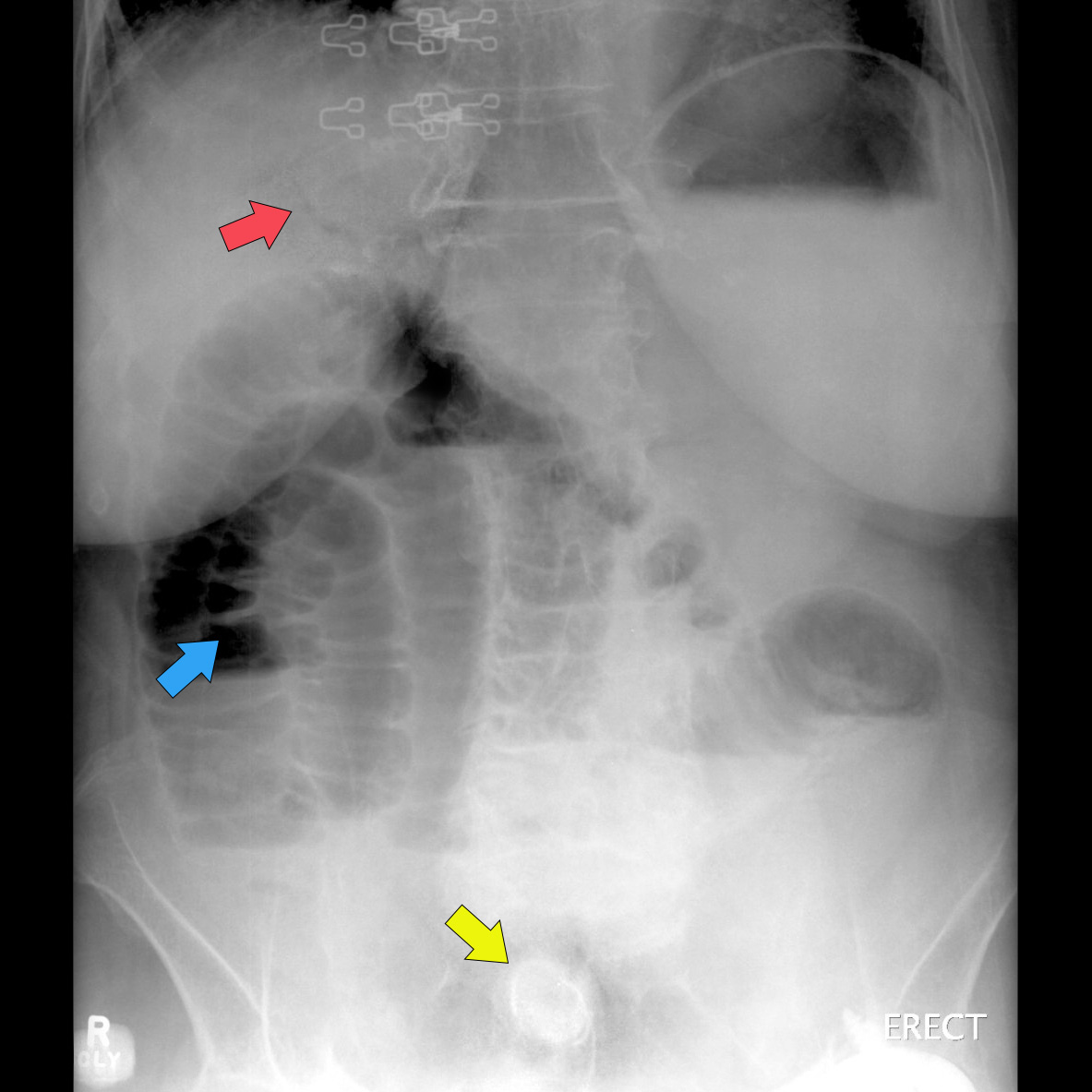
The condition was first described by the Danish physician Thomas Bartholin in 1654. A characteristic radiographic appearance was described by Dr. Leo Rigler (Rigler’s triad) in 1941:
- evidence of small bowel obstruction (such as dilated bowel loops);
- a gallstone not in the area of the normal gallbladder;
- air in the bile ducts
Below: An abdominal radiograph showing Rigler’s triad: air in the bile ducts (red arrow), dilated small bowel loops (blue arrow), and a calcified mass (gallstone) in the lower abdomen (yellow arrow).
Source: Gaillard, F. (2009). Gallstone ileus. Radiopaedia. https://radiopaedia.org/cases/gallstone-ileus