
Age/sex: 20 year old male
Size: 16.4 x 21.3 x 7.3 cm
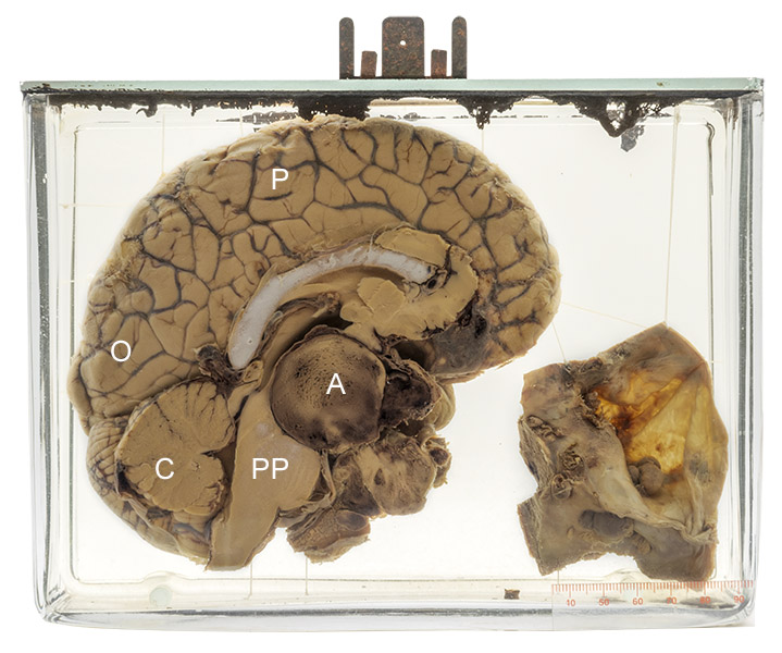
The specimen shows the brain cut in half in the mid-sagittal plane: cerebellum (C); parietal (P) and occipital (O) lobe cortex; pons (PP). A round, well circumscribed adenoma (A) is present in the region of the pituitary gland.
Pituitary adenoma
Tumors of the pituitary gland can be benign (about two thirds of cases) or locally invasive; rarely, they spread (metastasize) within the cranial cavity. They can result in a variety of signs and symptoms. Their size alone can cause headache or neurological findings such as visual disturbance by compression of adjacent tissue. Some also secrete hormones similar to those of the normal pituitary gland, causing conditions such as Cushing’s syndrome (ACTH) or acromegaly/gigantism (growth hormone). Some are a part of a multifocal tumor syndrome (e.g., multiple endocrine neoplasia, MEN1).
The normal pituitary gland is about 5 mm in diameter and is located in a depression in the sphenoid bone (sella turcica). It has both neural (posterior pituitary) and hormone secreting (anterior pituitary) components. Tumors that cause symptoms are relatively uncommon; however, small, usually asymptomatic tumors (“microadenomas”) are found in many individuals.
Below: A normal pituitary gland.
Source: Jomegat. (2006). Pituitary gland. Wikimedia Commons. https:// commons.wikimedia.org/wiki/File:Pituitary_gland.png
