
Age/sex: unknown
Size: 13.3 x 18.5 x 5.5 cm
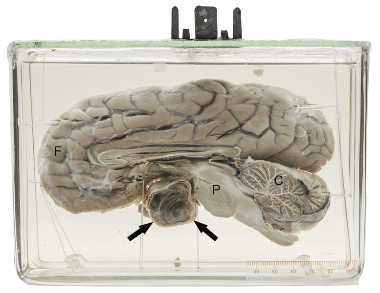
A mid-sagittal slice of the brain shows a well-circumscribed nodule (arrows) just anterior to the pons (P). Its dark color and laminated appearance suggest that it is composed of thrombus (clotted blood). The thin white tissue layer surrounding it is the arterial wall (the vessel has been cut in half).
(C = cerebellum; F = frontal lobe)
Cerebral aneurysms
An aneurysm is a localized dilatation of a vessel. It can affect any vessel in the body and range in size from mms to as much as 10 cm. There are many causes, including infection, immunologic disease (vasculitis), and atherosclerosis. Brain aneurysms are most often found at the branch points of a group of communicating vessels on its base called the Circle of Willis. They are believed to be related to a congenital weakening of a part of the wall which dilates over time because of blood pressure.
As the aneurysm’s diameter increases, the risk of its rupture also increases (similar to an inflated balloon). Most often, such rupture occurs in the subarachnoid space. The affected individual typically complains of headache, which can be severe. In many cases, it is followed by loss of consciousness and death as increasing bleeding causes brain herniation.
Thomas Willis (1621 – 1675) was an English physician who was particularly interested in the anatomy of the brain, nerves and muscles. He was responsible for describing the cranial nerves as well as the vascular arrangement at the base of the brain in his published Cerebri Anatome of 1664. He also introduced the word “neurology” to refer to the study of nervous disease.
Below: Base of brain showing the vascular Circle of Willis.
Source: Anatomist90. (2011). Circle of Willis. Wikimedia Commons. https:// commons.wikimedia.org/w/index.php?curid=17631847
