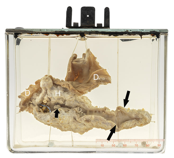
Age/sex: 52-year-old female
Size: 14 x 16 x 5.0 cm
The duodenum (D) is wrapped around the head of the pancreas (H). A blue rod can be seen in the pancreatic duct near the ampulla of Vater (the entry site of the pancreatic duct into the duodenum). Distally, the duct is dilated (large arrows) and the adjacent pancreatic tissue is markedly thinned (atrophic). The proximal duct contains several small stones (small arrow).
Chronic pancreatitis
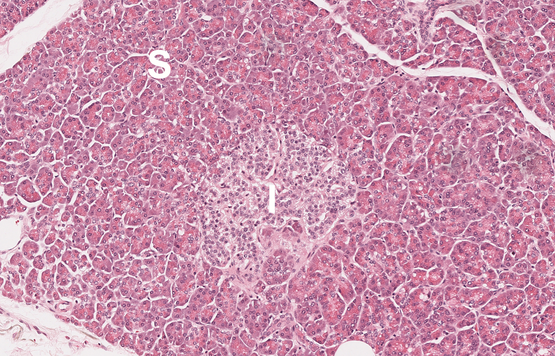
The pancreas has two major functions, one exocrine and the other endocrine. The first involves secretion of enzymes into the small intestine to aid in food digestion. The enzymes are made in serous cells, which secrete them via ducts that lead to the duodenum. They include trypsinogen (to digest proteins), amylase (for carbohydrates) and lipase (for fats). Pancreatic endocrine function is concerned primarily with control of glucose (sugar) metabolism via enzymes such as insulin and glucagon. These substances are made by cells located in small clusters known as Islets of Langerhans from which they are secreted directly into the blood, insulin causing a decrease in blood glucose and glucagon an increase.
Chronic pancreatitis has several causes, including obstruction of the main pancreatic duct by one or more stones (such as in this case) and direct injury of pancreatic cells by toxins such as alcohol. Clinically, it can be manifested by abnormalities in digestion (such as the presence of fat in the stool due to lipase deficiency) or in metabolism (such as diabetes secondary to loss of insulin).
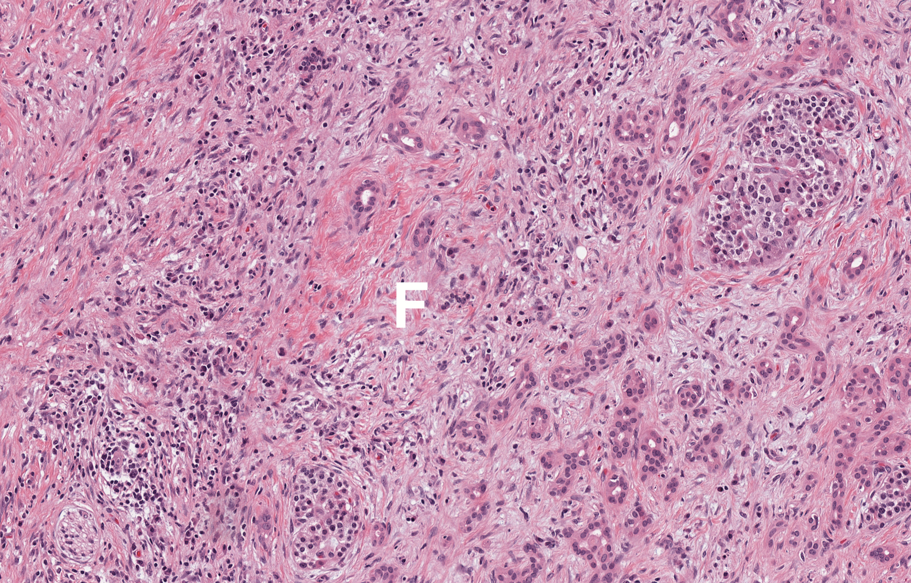
Below: Histologic appearance of normal pancreas (I- Islet of Langerhans; S- serous cells) and chronic pancreatitis (F- fibrosis; note loss of serous cells).
Source: Rajaram, A (2023). Normal pancreas and chronic pancreatitis.