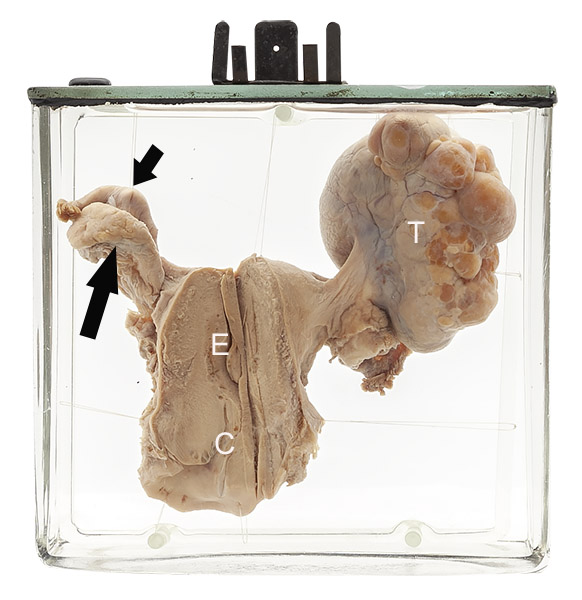
Age/sex: unknown
Size: 14.5 x 15.2 x 5.7 cm
The uterus has been opened to show the endometrial cavity (E) and endocervical canal (C). The left ovary (long arrow) and Fallopian tube (short arrow) are normal. A lobulated 8 cm tumor (T) is present where the right ovary should be.
Cystadenoma of ovary
Cystadenomas are the most common primary ovarian tumors, accounting for about 20 - 30% in most reviews. They may be serous (filled with clear fluid, as was likely the case in this specimen) or mucinous (containing mucoid secretions). The latter type can become very large, sometimes the size of a term baby!
Cystadenomas are almost always benign - i.e., they do not invade adjacent tissues or spread to other sites in the body. However, mucinous tumors particularly can be associated with small foci of cell proliferation on their surface, a finding that suggests the potential for an aggressive behaviour; such tumors are termed “borderline” cystadenomas.
Most tumors are discovered incidentally by finding a pelvic mass on physical examination or radiologic imaging. Larger ones may cause a feeling of pressure or aching in the pelvis and are sometimes associated with urinary frequency or constipation because of compression of the bladder or colon. Rarely, a tumor twists, leading to occlusion of its feeding and/or draining blood vessels; this can be painful and is sometimes followed by cyst necrosis and rupture.
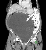
Below: CT image showing a large, mucinous ovarian cystadenoma (arrow).
Source: Ashraf, A. (2021). Ovarian mucinous cystadenoma. Radiopaedia. https://radiopaedia.org/cases/ovarian-mucinous-cystadenoma-12?lang=us