Age/sex: 37-year-old female
Size: 16.3 x 11.5 x 5.2 cm
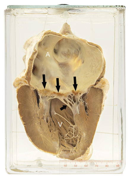
The specimen consists of a heart with a moderately dilated left atrium (A) and a normal left ventricle (V). The mitral valve leaflets appear thickened and the chordae tendineae (short arrow) are thickened and fused (compare with those in Specimen 40). This appearance is consistent with rheumatic mitral valve stenosis. A row of finely granular, tan-colored vegetations (long arrows) is present on the upper valve surface.
Non-bacterial thrombotic endocarditis
Although it is not possible to be certain without microscopic examination, the small and uniform size of the vegetations in this specimen and the absence of injury to the underlying valve leaflets suggest that these growths are not the result of infection.
This condition is the result of a combination of the normal mildly turbulent blood flow across the mitral valve and a hypercoagulable state. The latter has many causes, and can be seen normally in pregnancy or in disease such as antiphospholipid syndrome and some forms of cancer. The vegetation consists only of thrombus, unlike infectious endocarditis in which neutrophils and bacterial clusters are also present and are usually associated with damage to the adjacent valve leaflet.
The condition is usually of no clinical importance. However, fragments of the vegetations may be dislodged and spread (embolize) elsewhere in the body where they can cause serious disease, such as stroke (cerebral infarction).
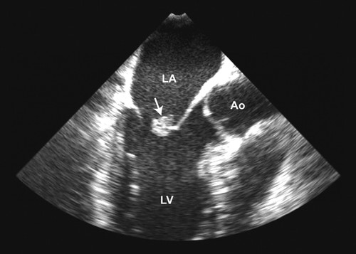
Below: An image from a cardiac ultrasound showing a small mass on the mitral valve (arrow) representing non-bacterial thrombotic endocarditis in a patient with antiphospholipid syndrome (LA = left atrium; LV = left ventricle; Ao = aorta).
Source: Figure 2. Mass on the mitral valve. From “Unusual case of nonbacterial thrombotic endocarditis attributable to primary antiphospholipid syndrome,” by KL Vinales, RS Gopalan, LA Lanza, et al. (2010). Circulation 122(12), 459-460.