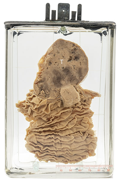
Age/sex: 51-year-old male
Size: 16.4 x 11.7 x 5.4 cm
The specimen consists of a segment of jejunum with prominent mucosal folds (valvulae conniventes). Attached to the serosa is a well-circumscribed gray tumor (T) measuring about 7 cm in diameter.
Leiomyoma: Gastrointestinal stromal tumor
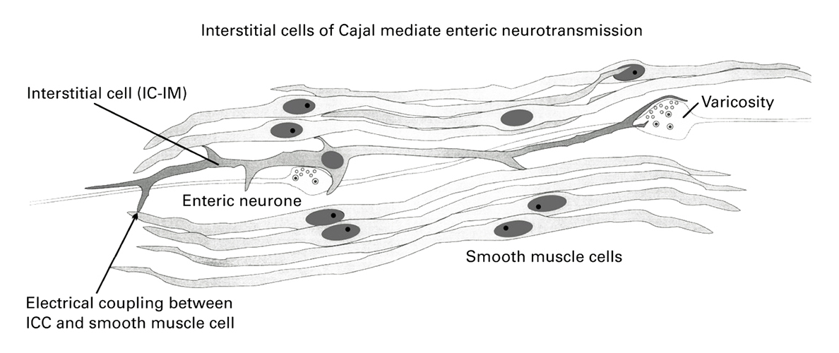
Leiomyomas are benign neoplasms (tumors) of smooth muscle. They can be found anywhere in the body in which smooth muscle is normally present, including the bowel. In women, they are by far most common in the uterus (see Specimen 22). Histologically, they are composed of regularly shaped cells that have an elongated (spindled) appearance similar to the normal smooth muscle cell. Nowadays, most intestinal tumors that show this histologic pattern are called gastrointestinal stromal tumors (GISTs). Rather than originating in smooth muscle, they are thought to arise from special cells in the gastrointestinal tract called interstitial cells of Cajal that cause its smooth muscle to contract and move food distally.
GISTs are often small and not associated with symptoms. However, they can become large enough to cause intestinal obstruction, erode the overlying mucosa and cause bleeding, or serve as a focus (leading edge) for bowel intussusception (see Specimen 51). Although most GISTs are benign, they can invade adjacent tissue and spread to other parts of the body (metastasize). Thus, if symptoms do occur or if they are larger than 2 cm, they are usually removed surgically.
Below: Diagram of a portion of small bowel wall showing an interstitial cell coupled with several smooth muscle cells.
Source: Fig. 3 Ward SM Interstitial cells of Cajal in enteric neurotransmission Gut 2000;47:iv40-iv43. British Society of Gastroenterology.