
Age/sex: 47-year-old female
Size: 16.7 x 17.2 x 6.5 cm
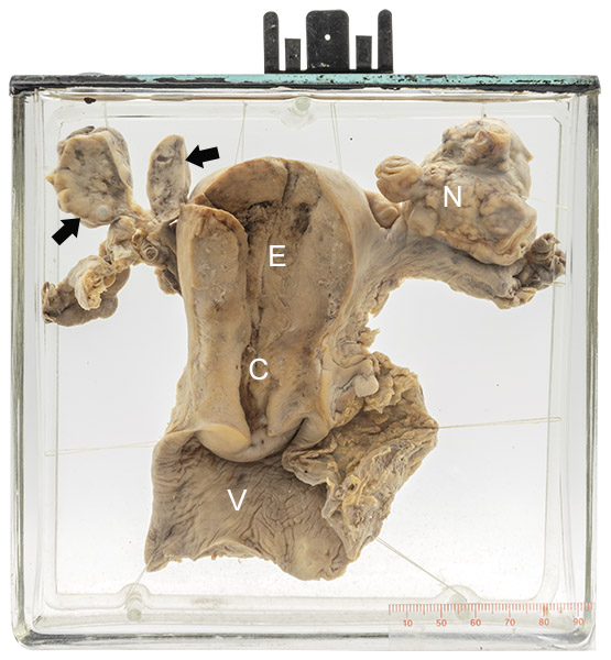
The uterus has been opened to show the endometrial cavity (E) and endocervical canal (C). The upper portion of the vagina (V) is attached. Both ovaries are enlarged and nodular (N) (compare with a normal ovary in Specimen 19). The cut surface of two of the nodules (arrows) shows them to have a white appearance consistent with carcinoma. The patient was diagnosed with breast carcinoma one month before death.
Metastatic carcinoma to ovary
Metastasis – spread of tumor tissue from one place in the body to another – is common in cancer. The most frequent sites of such spread are lymph nodes near the region of the primary tumor (for example the axilla in breast carcinoma), the lung, liver, and bone. However, any organ or tissue, including the ovary, can be affected.
The condition is not uncommon in the ovary: about 15 - 20% of all ovarian cancers are metastases from somewhere else. The stomach, colon and breast are the most common sites of origin. Although such tumors are most often part of widespread metastatic disease, in some individuals the ovary is the only site. When such cancer has a signet ring cell appearance on microscopic examination, it is sometimes termed a “Krukenberg tumor”.
Friedrich Krukenberg (1871 – 1946) was a German physician who reported in 1896 what he believed was a new form of primary ovarian cancer that he called fibrosarcoma ovarii mucocellulare carcinomatodes. (In fact, the same tumor had been described by James Paget in 1854.) Although Krukenberg was also incorrect in his belief that the tumor originated in the ovary – it was shown to be metastatic, most often from the stomach, 6 years later - his name has nevertheless become associated with it.
Below: Histology of a carcinoma composed of signet ring cells (arrow) consistent with a Krukenberg tumor. The pale blue appearance of the cytoplasm is the result of mucous that distends the cell (representing a finger); the compressed cell nucleus next to it represents the ring.
Source: Rajaram, A. (2023). Signet ring adenocarcinoma
![]()